

Researchers at Northwestern University have developed the first physics-based measure to predict whether a person might one day suffer from an aortic aneurysm, a fatal condition that often causes no symptoms until it ruptures.
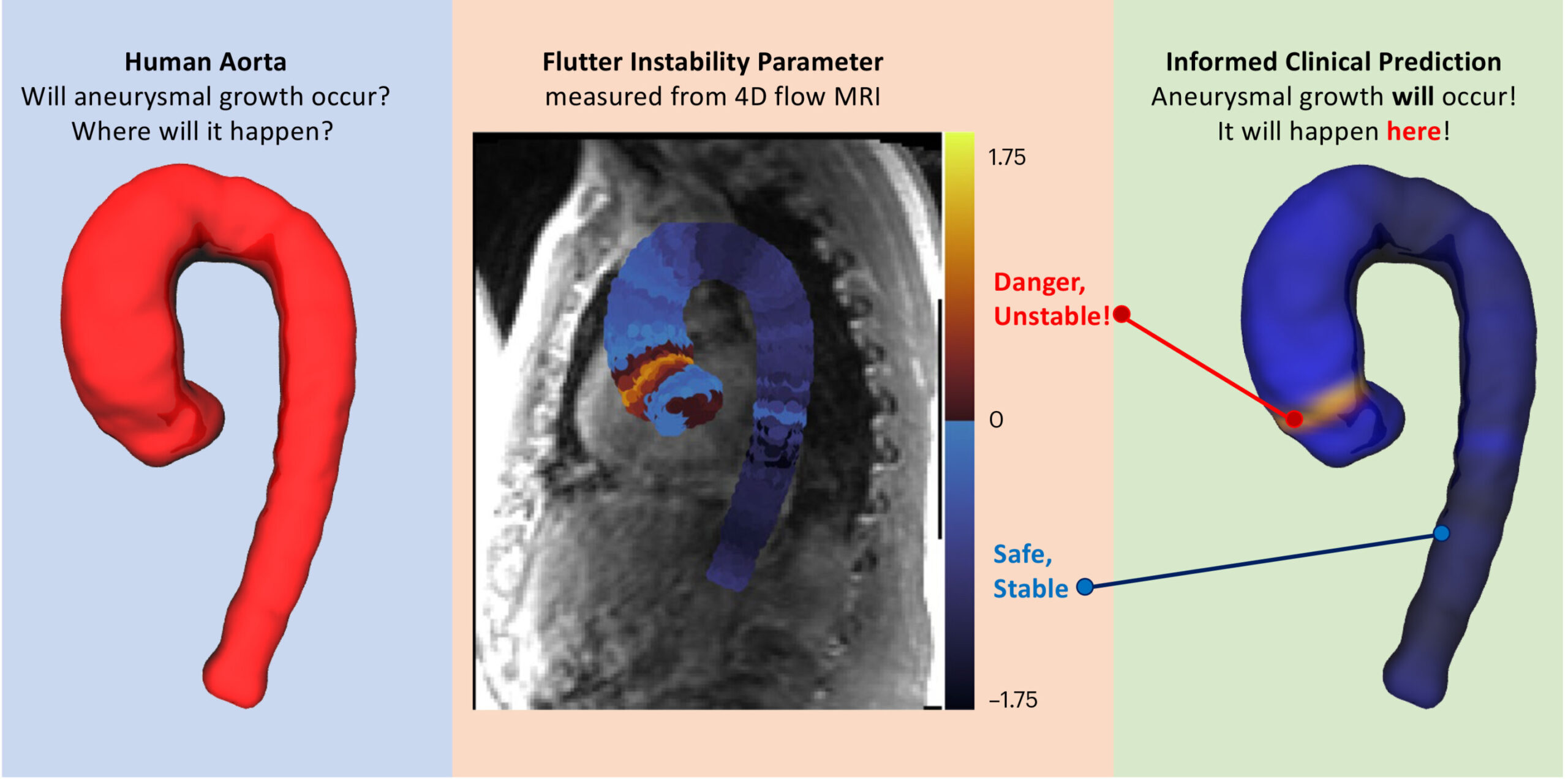
In the new study, researchers predicted abnormal aortic growth by measuring subtle “beats” in a patient’s blood vessels. As blood flows through the aorta, the vessel wall can flutter, much like a banner waves in the breeze. While stable flow predicts normal, natural growth, unstable flutter is strongly predictive of future abnormal growth and potential rupture, the researchers found.
Called the “float instability parameter” (FIP), the new measurement predicts future aneurysm with 98% accuracy on average three years after the first FIP measurement. To calculate a personalized PIF, patients only need a single 4D flow magnetic resonance imaging (MRI) scan.
Using the predictive and clinically measurable measure, doctors could prescribe drugs to high-risk patients to intervene and potentially prevent the aorta from swelling to a dangerous size.
The research was published this week (December 11) in the journal Natural biomedical engineering.
“Aortic aneurysms are colloquially called ‘silent killers’ because they often go unnoticed until catastrophic dissection or rupture occurs,” said Neelesh A. Patankar of Northwestern, lead author of the study. . “The fundamental physics behind aneurysms is unknown. As a result, there are no clinically approved protocols for predicting them. We have now demonstrated the effectiveness of a physics-based measure that helps predict future growth “This could be transformational in predicting cardiac pathologies.”
An expert in fluid dynamics, Patankar is a professor of mechanical engineering at Northwestern’s McCormick School of Engineering. He co-led the study with Dr. Tom Zhao, who specializes in first principles of biomechanics.
A growing danger
An aortic aneurysm occurs when the aorta (the largest artery in the human body) swells to a size greater than 1.5 times its original size. As it grows, the wall of the aorta weakens. Eventually, the wall becomes so weak that it can no longer withstand the pressure of the blood passing through it, causing the aorta to rupture. Although rare, an aortic rupture is usually unpredictable and almost always fatal.
Several famous people have died from aortic aneurysms, including Grant Wahl, a sports journalist who died suddenly a year ago during the 2022 FIFA World Cup. Other celebrity deaths include John Ritter, Lucille Ball and Albert Einstein.
“Most people don’t realize they have an aneurysm unless it’s detected accidentally during a CT scan for an unrelated problem,” Patankar said. “If doctors detect it, they may suggest lifestyle changes or prescribe medications to lower blood pressure, heart rate and cholesterol. If this is not detected, it may rupture, which is a immediate catastrophic event.”
“If it ruptures when the person is outside a hospital, the mortality rate is close to 100 percent,” Zhao added. “The blood supply to the body stops, so essential organs like the brain can no longer function.”
Remove uncertainties
For current standards of care, doctors estimate the risk of rupture based on risk factors (such as age or smoking history) and the size of the aorta. To monitor a growing aorta, doctors follow it with regular imaging tests. If the aorta begins to grow too quickly or become too large, the patient will often undergo a surgical graft to strengthen the vessel wall, an invasive procedure that carries its own risks.
“Our collective lack of understanding makes it difficult to track aneurysm progression,” Zhao said. “Doctors should regularly track the size of an aneurysm by viewing its location every one to five years, depending on how quickly it grew previously and whether the patient has any associated diseases. During this period waiting, an aneurysm can burst fatally.
To eliminate the uncertainties in predicting future aneurysms, Patankar, Zhao and their collaborators sought to grasp the fundamental physics underlying the problem. Through extensive mathematical work and analysis, they discovered that problems arise when the floating vessel wall changes from stable to unstable. This instability causes or signals an aneurysm.
“Float is a mechanical signature of future growth,” Patankar said.
Capturing the underlying physics
To quantify the transition from stability to instability, the researchers combined blood pressure, aortic size, aortic wall stiffness, wall shear stress, and pulse rate. The resulting number (or FIP) characterizes the exact interaction between blood pressure and wall stiffness that ultimately triggers floating instability.
“Doctors knew these factors — blood pressure, heartbeat rate and aorta size — were involved, but they didn’t know how to quantify it,” Patankar said. “It turns out that the combination of these factors is what’s important. A patient may have an unstable wall but a normal-sized aorta, so their doctor won’t even realize there’s a problem. ”
Surprisingly, the researchers found that instability tends to occur when the wall is more flexible. This finding directly contradicts the common knowledge that aortic stiffness is a sign of disease.
“We show that the less stiff it is, the more risk the patient has for future growth and rupture,” Zhao said. “Indeed, once the aorta reaches a certain size, the body tries to stiffen it to apparently protect it from future growth. But those that continue to grow are less rigid. The aorta will float if the wall is more flexible.”
Validate the metric
To test this new measure, researchers examined 4D flow MRI data from 117 patients who underwent cardiac imaging to monitor heart disease and 100 healthy volunteers. Based on this MRI, the researchers assigned each patient a personalized PIF. In this metric, zero marks the threshold between stable and unstable.
For patients with a PIF less than zero, their aorta was unlikely to experience abnormal growth. The researchers predicted, however, that patients with a PIF greater than zero would experience abnormal growth and future rupture.
“By establishing the prognostic value of this quantitative measure for 4D cardiovascular MRI flow, we can significantly improve the value of imaging offered as a standard of care for patients with aneurysms,” said Dr. Ethan Johnson, co -first author of the study and postdoctoral researcher. in cardiovascular imaging from the Feinberg School of Medicine at Northwestern University.
When the researchers compared these predictions to follow-up MRIs or doctors’ diagnoses, they found that their predictions were accurate 98% of the time. Although the FIP predicted future growth on average three years after the initial MRI (when the FIP was calculated), the researchers say this measure could even provide a more granular view of heart health on a daily or monthly basis. .
“The one to eight year period is the time frame that our clinical data falls into,” Zhao said. “Not the total time interval over which FIP is necessarily effective.”
Next, Patankar, Zhao and their team plan to explore whether FIP can provide clues about how other heart diseases develop. They are also studying whether patient-specific PIF can indicate which prevention methods are most effective in stopping aneurysm progression.
The research is titled “Blood Wall Floating Instability as a Physiomarker of Progression of Thoracic Aortic Aneurysms.”
More information:
Tom Y. Zhao et al, Floating blood wall instability as a physiomarker of progression of thoracic aortic aneurysms, Natural biomedical engineering (2023). DOI: 10.1038/s41551-023-01130-1
Provided by Northwestern University
Quote: Unstable ‘float’ predicts aortic aneurysm with 98% accuracy (December 16, 2023) retrieved December 17, 2023 from
This document is subject to copyright. Apart from fair use for private study or research purposes, no part may be reproduced without written permission. The content is provided for information only.
