by Kunmo Koo, Xiaobing Hu and Vinayak P. Dravid
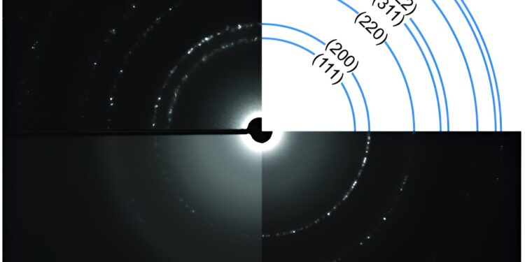
Comparisons of diffraction data with different support structures. Credit: Kunmo Koo
When someone suggests the word “enlarge”, they are referring either to bringing distant objects closer together or to enlarging small objects to a tangible scale. There is no doubt that the power of magnifying instruments, regardless of their scale and direction, can lead to progress in the scientific field. Since its launch in 2021, the James Webb Space Telescope (JWST) has embarked on a mission to collect unprecedented data about the deep universe, with the aim of expanding our understanding of the early universe and the cycle life of celestial bodies.
The appropriate analogy for JWST in the atomic world is the aberration-corrected electron microscope (ACEM). By exploiting a highly coherent electron as well as an aberration corrector, the microscope excels in resolving subatomic features, enabling comprehensive exploration of the structure-functional relationship in materials. As an essential element for nanoworld navigators, modern ACEM can provide invaluable information that remains irreplaceable by other characterization methods.
The contradiction comes from the dual nature of high energy electrons. The wave property of the electron allows high-resolution imaging, while the particle property makes collisions inevitable. When the particle moves through the gas at ambient pressure, its mean free path (the distance it can travel before substantially changing its original direction or energy) is limited to only about 100 nm.
Ballistic collisions change the direction of the electron or deplete its energy, significantly hampering the performance of electron optics. To avoid these collisions, the microscope column is usually kept in ultra-high vacuum conditions, which are at least 10ten times thinner than ambient air.
The nature of ACEM limits its applicability to static, thin and solid samples. However, materials encompass various states of matter beyond solids, including liquids, gases, and plasmas. To observe reactions at the nanoscale, it is essential to encapsulate the fluidic media involved in a sealed nanoreactor, thereby preventing their dissipation. The use of silicon nitride microelectromechanical systems (MEMS) technology addresses these particular needs, allowing researchers to explore reactions at the nanoscale.
Electron microscopy image of ultra-thin beehive-inspired silicon nitride. Credit: Kunmo Koo
The silicon nitride film, serving as the encapsulation membrane, can be conveniently produced with a thickness on the order of tens of nanometers using a chemical vapor deposition process. These films exhibit reasonable resilience to mechanical shock, particularly when over a certain thickness, although there is a trade-off relationship with electronic transparency.
Like an aquarium with a wall of glass several feet thick, which may be sturdy enough to hold a large amount of water, maximizing visibility through the glass becomes a challenge. Therefore, the engineering of the “wall” is crucial to ensure optimal visibility in the aquariums and in the ACEM liquid tank.
To meet this challenge, we take inspiration from the beehive, a structure that resists high mechanical stresses while using a minimum of material. Our solution is to create a space-filling hexagonal support system using heavily doped silicon under ultra-thin silicon nitride, which is only 1/5th the thickness of the conventional method.
The beehive-shaped structure maximizes the opening for observing reactions and provides optimal resistance under mechanical stress. With this ultra-thin breakthrough, the membrane can be thinned down to a single-digit nanoscale, or about 1/10,000th the thickness of a human hair, without experiencing rupture or leaking under the microscope.
The transparency of the ultrathin membrane enables fluid mapping with sub-nanometer spatial resolution and significant suppression of unwanted electron scattering, a capability not achievable with conventional cladding materials. This breakthrough enables gas-phase sensitivity to the point of detecting a handful of gas molecules inside the transmission electron microscope (TEM). This level of sensitivity makes it possible to capture reactions occurring at the gas-solid interface with temporal resolution on the microsecond scale.
As an illustrative example, we visualize the insertion of hydrogen atoms into metallic palladium under ambient temperature and pressure conditions. This technology holds immense potential for the development and study of nanocatalysts for gas-phase carbon capture, as well as for energy materials such as fuel cells and metal-air batteries, thereby providing information to the atomic scale. Our work is published in the journal Scientists progress.
Although operating at a different scale and scope, we draw parallels between this development and the revolutionary capabilities of the James Webb Space Telescope (JWST), which provides unprecedented images and data that challenge cosmological theories. Furthermore, we propose that this innovative strategy for designing ultrathin membrane microchips can be extended to various applications in which thin membranes serve as encapsulation and/or support materials, with implications extending beyond the field nanosciences.
This story is part of Science X Dialog, where researchers can report the results of their published research articles. Visit this page for more information about ScienceX Dialog and how to get involved.
More information:
Kunmo Koo et al, Ultra-thin silicon nitride microchip for in situ/operando microscopy with high spatial resolution and spectral visibility, Scientists progress (2024). DOI: 10.1126/sciadv.adj6417
Dr. Kunmo Koo is a research associate at the NUANCE Center. Dr. Xiaobing Hu is a Research Associate Professor in the Department of Materials Science and Engineering and Head of TEM Facilities at the NUANCE Center. Dr. Vinayak P. Dravid is the Abraham Harris Professor of Materials Science and Engineering and founding director of the NUANCE Center.
Quote: Ultrathin membranes to discover the problem at the atomic scale under operando conditions (January 30, 2024) retrieved on January 30, 2024 from
This document is subject to copyright. Except for fair use for private study or research purposes, no part may be reproduced without written permission. The content is provided for information only.


