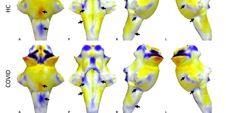
3D projections of QSM χ maps on rendered brainstem ROI extracted from FreeSurfer segmentation for the healthy control group and the COVID group. The COVID group shows increased χ in the brainstem, particularly in the medulla and Pons (black arrows). Credit: University of Cambridge
Damage to the brainstem – the brain’s “control center” – causes the lasting physical and psychiatric effects of severe COVID-19 infection, a study suggests.
Using ultra-high resolution scanners capable of observing the living brain in detail, researchers from the Universities of Cambridge and Oxford have been able to observe the damaging effects that COVID-19 can have on the brain.
The study team scanned the brains of 30 people who had been admitted to hospital with severe COVID-19 early in the pandemic, before vaccines were available. Researchers have found that COVID-19 infection damages the brainstem region associated with shortness of breath, fatigue and anxiety.
The powerful MRI scanners used for the study, known as 7 Tesla or 7T scanners, can measure inflammation in the brain. Their results, published in the journal Brainwill help scientists and clinicians understand the long-term effects of COVID-19 on the brain and the rest of the body. Although the study was launched before the long-term effects of COVID were recognized, it will help better understand this disease.
The brainstem, which connects the brain to the spinal cord, is the control center for many vital functions and fundamental reflexes. Clusters of nerve cells in the brainstem, called nuclei, are responsible for regulating and processing essential bodily functions such as breathing, heart rate, pain, and blood pressure.
“Events that occur in and around the brainstem are essential to quality of life, but it was impossible to scan the inflammation of brainstem nuclei in living people, due to their small size and difficult position .” said first author Dr. Catarina Rua, from the Department of Clinical Neurosciences. “Usually, scientists only get a good look at the brainstem during post-mortem examinations.”
“The brainstem is the essential junction box between our consciousness and what’s happening in our body,” said Professor James Rowe, also from the Department of Clinical Neuroscience, who co-led the research. “The ability to see and understand how the brainstem changes in response to COVID-19 will help explain and treat long-term effects more effectively.”
Early in the COVID-19 pandemic, before effective vaccines were available, postmortem studies of patients who died from severe COVID-19 infections showed changes in their brainstems, including inflammation. Many of these changes were thought to result from a post-infection immune response, rather than a direct invasion of the brain by the virus.
“People who were very sick early in the pandemic showed lasting brain changes, likely caused by an immune response to the virus. But measuring that immune response is difficult in living people,” Rowe said. “Normal hospital-style MRI scanners cannot see the inside of the brain in the kind of chemical and physical detail we need.”
“But with the 7T scanners, we can now measure these details. Active immune cells interfere with the ultra-high magnetic field, so we are able to detect their behavior,” Rua said. “Cambridge was special because we were able to scan even the sickest and most contagious patients, early in the pandemic.”
Many patients admitted to the hospital early in the pandemic reported fatigue, shortness of breath and chest pain as troubling long-term symptoms. The researchers hypothesized that these symptoms were in part the result of damage to key nuclei in the brainstem, damage that persists long after COVID-19 infection ends.
The researchers found that several regions of the brainstem, particularly the medulla oblongata, pons and midbrain, showed abnormalities consistent with a neuroinflammatory response. The abnormalities appeared several weeks after hospital admission and in regions of the brain responsible for controlling breathing.
“The fact that we see abnormalities in parts of the brain associated with breathing strongly suggests that the long-lasting symptoms are an effect of brainstem inflammation following COVID-19 infection,” Rua said. “These effects are additive to the effects of age and sex, and are more pronounced in those who have had severe COVID-19.”
In addition to the physical effects of COVID-19, the 7T scans have highlighted some of the psychiatric effects of the disease. The brainstem monitors shortness of breath, as well as fatigue and anxiety. “Mental health is intimately linked to brain health, and patients with the strongest immune response also had higher levels of depression and anxiety,” Rowe said.
“Changes in the brainstem caused by COVID-19 infection could also lead to poor mental health outcomes, due to the close connection between physical and mental health. »
The researchers say the findings could help understand other conditions associated with brainstem inflammation, such as MS and dementia. The 7T scanners could also be used to monitor the effectiveness of different treatments for brain diseases.
“This was an incredible collaboration, right at the height of the pandemic, when testing was very difficult, and I was amazed at how well the 7T scanners worked,” Rua said. “I was really impressed by how, in the heat of the moment, the collaboration between many different researchers happened so effectively.”
More information:
Catarina Rua et al, Quantitative mapping of 7T susceptibility in COVID-19: brainstem effects and outcome associations, Brain (2024). DOI: 10.1093/brain/awae215
Brain
Provided by the University of Cambridge
Quote: Ultra-powerful MRIs show damage to brain’s ‘control center’ causes lasting COVID-19 symptoms (October 7, 2024) retrieved October 7, 2024 from
This document is subject to copyright. Except for fair use for private study or research purposes, no part may be reproduced without written permission. The content is provided for informational purposes only.


