

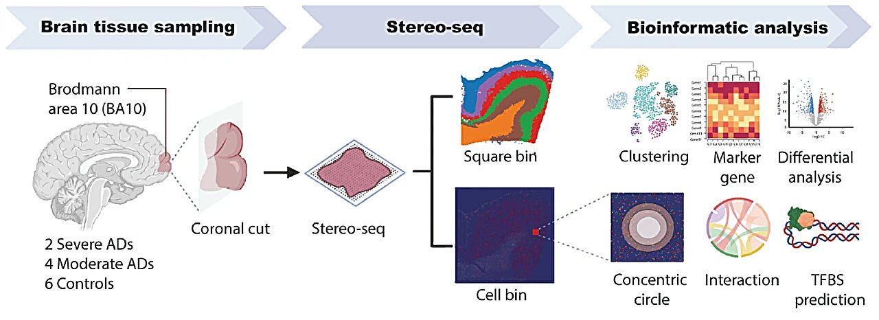
The study analysis pipeline, with selected figures created using Biorender. Credit: Nature communications (2025). DOI: 10.1038 / S41467-024-54715-Y
Researchers at the University of Tulane created a first subcellular map of his kind of a brain area commonly affected by Alzheimer’s disease, a key step towards disentangling mysteries of the way in which degenerative brain disease develops.
The study, published in Nature communicationsThe genetic mechanisms that cause the loss of brain cells that allow the disease to progress and identify a key protein as a potential treatment target.
More than 55 million people worldwide suffer from dementia, with Alzheimer’s disease for 60% to 70% of these cases. Despite the prevalence, little is known about its cause and existing drugs can only temporarily facilitate symptoms, not preventing the disease from progressing.
“The human brain is the most complex organ of the human body and the mechanism of many diseases like Alzheimer’s is elusive,” said the main author Hui Shen, associate director of the Center for Biomedical Informatics & Genomics to the Tulane University School of Medicine. “Using the spatial transcriptomic, we were able to create a card from a part of the prefrontal cortex with a unique resolution to try to understand the underlying factors of Alzheimer.”
The researchers used stereo sequencing to examine a small section of the prefrontal cortex – the region responsible for decision -making and emotional control – in six brains at different stages of Alzheimer.
This technology allowed them to “map” the brain tissue almost 250 times the resolution of older tools, essentially zooming in revealing genetic interactions within a single cell and how people travel as the disease progresses.
The study revealed that genetic modules responsible for protecting neurons weaken or disappear in Alzheimer’s patients, allowing harmful proteins linked to the disease to build and damage cells.
The researchers identified a protein, ZNF460, as crucial for the neuroprotective processes of these modules and as a potential treatment target.
“The most important thing is that we have identified several interesting interactions at the molecular level that work to protect neurons under stress, and these interactions have disappeared in Alzheimer’s patients,” said the main author Yun Gong, an instructor for the Center for Biomedical Informatics & Genomics at the Tulane University School of Medicine. “If we can find a way to target ZNF460 in a way that maintains these modules, then we could be able to inhibit the progress of Alzheimer’s.”
In another surprising discovery, the study revealed that the layer structure of the brain disappears as the disease progresses, a phenomenon which, according to Gong, had not been observed before “.
In the future, Shen and Gong declared that they hoped to seek research on the ZNF460 and check if its absence alone can be linked to the beginning of Alzheimer’s.
“It’s just a step towards understanding the pathophysiology of Alzheimer’s disease,” said Shen. “Different areas of the brain can respond differently to the development of the disease, so we must continue to work to examine other regions and create the most complete image as possible.”
More information:
Yun Gong et al, Stereo-Seq of the prefrontal cortex in aging and Alzheimer’s disease, Nature communications (2025). DOI: 10.1038 / S41467-024-54715-Y
Supplied by Tulane University
Quote: The new brain cartography technique highlights the development of Alzheimer’s (2025, May 5) recovered on May 5, 2025
This document is subject to copyright. In addition to any fair program for private or research purposes, no part can be reproduced without written authorization. The content is provided only for information purposes.
