

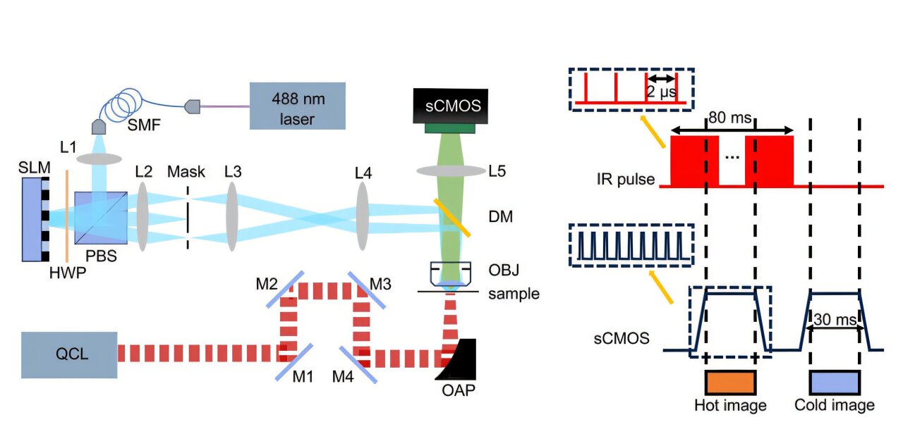
Simip allows high -resolution images rich in chemical and spatial information. A quantum cascade laser (QCL) excites molecular vibrations while a spatial light modulator (SLM) generates striped light patterns which are projected on the sample. A Scientific CMOS (SCMOS) camera captures modulated fluorescence signals, which are treated using Hesse Dédévolution and Clear Devolution algorithms to generate high -resolution chemical and structural images. The subtraction of the hot image of the cold image gives the hybrid similar image. Credit: Advanced photonics (2025). DOI: 10.1117 / 1.AP.7.3.036003
Today’s super-resolution microscopes have made it possible to observe the nanometric world with unprecedented details. However, they require fluorescent labels, which reveal structural details but provide little chemical information on the samples studied.
This drawback has led to the development of vibratory imaging techniques, which can identify molecules according to their unique chemical links without modifying the sample. These methods detect physical changes in samples when they absorb medium infrared light (MIR), such as offsets of the refractive index caused by heat absorption or acoustic signals induced by temperature. And yet, existing methods often fight with low signal levels, which makes it difficult to make both high resolution (how finely details can be seen) and a strong chemical contrast (how molecules can be distinguished).
As indicated in Advanced photonicsA newly developed technique, structured illumination of photothermical microscopy (Simip), now addresses this limitation with a better resolution than conventional microscopy.
Developed by researchers from the University of Zhejiang, in China, led by Professor Delong Zhang, the new technique represents a significant progression of vibratory imagery, opening up new possibilities for chemical and biological analysis on a nanometric scale.
Zhang Note: “Simip microscopy incorporates the principles of structured microscopy with lighting with photothermic detection in a median medium. Middle infrared photodetection provides chemical specificity, while structured lighting microscopy improves the spatial resolution of the sample.”
The system consists of a quantum cascade laser (QCL) which excites specific molecular bonds, causing localized heating which reduces the brightness of the adjacent fluorescent molecules. Simultaneously, a SIM system composed of a continuous wave laser of 488 Nm and a spatial light modulator (SLM) generates striped light patterns which are projected on the sample with different angles.
These models create Moiré fringes, coding for high frequency details not resolved before in detectable low frequency signals which are captured by a scientific CMOS camera (SCMS). By comparing the images taken with and without vibrational absorption, Simip reconstructs high resolution images which are rich in chemical and spatial information.
The team applied the Sim Hesse and the sparse deconvolution algorithms to reach a higher spatial resolution, up to ∼60 Nm, with an imaging speed of more than 24 images per second, exceeding the conventional mir -mirm photatormic imagery.
To validate the precision of Simip, the researchers tested it on polymethyl methacrylate balls of 200 Nm incorporated with thermosensible fluorescent dyes. By sweeping the QCL through the 1,420–1,778 cm-1 The beach, Simip has successfully reconstructed vibrational spectra, closely corresponding to the results of the infrared spectroscopy (FTIR) of Fourier Transform (FTIR).
In terms of resolution, Simip has achieved an improvement of 1.5 times compared to the conventional mir-mit photothermical imagery, with a complete semi-maximum width (FWHM) of 335 Nm against 444 Nm in standard methods. In addition, he was able to distinguish the beads of polystyrene and methacrylate polymethyl in sub-dimming aggregates, which was impossible with standard fluorescence microscopy.
An additional advantage of Simip is its ability to detect autofluorescence – the natural fluorescence emitted by certain biological molecules. This can be done by passing from SIM with a large field to Punctual Balayage for a structured autoflorescence excitation or using a shorter wave beam for a large -field photoothermal detection method to improve compatibility with existing optical configurations.
By integrating the SIM with the MIP, Simip performs chemical imaging at high speed and super-resolution beyond the diffraction limit. This method opens up new possibilities of observations in materials science, biomedical research and chemical analysis. For example, researchers plan to use Simip to detect metabolites with small molecule and analyze their interactions with cellular structures.
The team now plans to improve the temporal synchronization of Simip to further improve imaging and precision speed, as well as exploring temperature -sensitive dyes to increase sensitivity. With minimum material changes to existing SIM systems, Simip is ready for adoption in laboratories around the world.
More information:
Pengcheng Fu et al, breaking the diffraction limit of molecular imaging by structured illumination microscopy medium infrared photothermics, Advanced photonics (2025). DOI: 10.1117 / 1.AP.7.3.036003
Quote: The microscopy method breaks barriers in chemical imaging on a nanometric scale (2025, April 14) recovered on April 15, 2025 from
This document is subject to copyright. In addition to any fair program for private or research purposes, no part can be reproduced without written authorization. The content is provided only for information purposes.
