
‘Cancer-Cooling’ Protein Puts Bowel Cancer on Ice
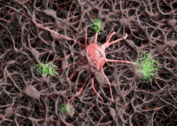
An immune system protein can be manipulated to help beat bowel cancer, according to new research from the Australian National ...

An immune system protein can be manipulated to help beat bowel cancer, according to new research from the Australian National ...

The human immune system constantly fends off a wide range of invaders, a feat that requires a wide array of ...
More than 700 million people have been infected and nearly seven million have died, making SARS-CoV-2 the most devastating pandemic ...
A team of scientists from the U.S. Department of Energy's (DOE) Brookhaven National Laboratory and Columbia University have demonstrated a ...
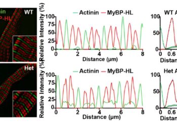
Investigators led by Elizabeth McNally, M.D., Ph.D., Elizabeth J. Ward Professor of Genetic Medicine and director of the Center for ...
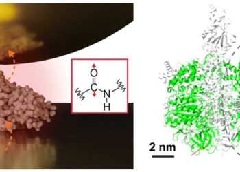
An interdisciplinary research team, led by Assistant Professor Jun Nishida and Associate Professor Takashi Kumagai of the Institute of Molecular ...
An international collaborative study recently discovered that variants in the PPFIA3 gene cause a previously unknown syndromic neurodevelopmental disorder. The ...

A pair of proteins, YAP and TAZ, have been identified as drivers of bone development in the uterus and may ...

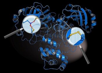
A team of researchers has developed an innovative method to engineer fully α complex proteins, characterized by their non-uniformly arranged ...

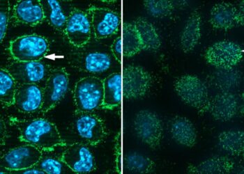
Just as healthy organs are essential to our well-being, healthy organelles are essential to the proper functioning of the cell. ...