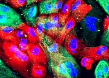
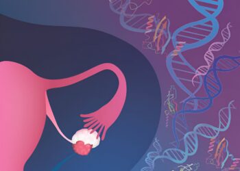
Scientists make COVID receptor protein in mouse cells
The scientists used fluorescence microscopy to visualize human ACE2 proteins expressed on mouse cells after incubating the cells with fluorescently ...
Structured illumination microscopy of atrial cardiomyocytes isolated from wild-type (A) and heterozygous Mybphl-null mice (B) reveals localization of MyBP-HL (red) ...
Single-protein infrared vibrational spectroscopy (left) Schematic of near-field infrared spectroscopy measuring a single protein. (Right) The structure of the F1-ATPase ...
Credit: Pixabay/CC0 Public domain An international collaborative study recently discovered that variants in the PPFIA3 gene cause a previously unknown ...
Graphical summary. Credit: Development cell (2023). DOI: 10.1016/j.devcel.2023.11.029 A pair of proteins, YAP and TAZ, have been identified as drivers ...
Credit: Nature Structural and molecular biology (2024) A team of researchers has developed an innovative method to engineer fully α ...
Overview: Mitochondrial and lysosomal stress stimulates nuclear translocation of TFEB, followed by increased expression of HKDC1. HKDC1 stabilizes PINK1 through ...
Tenm3 expression in the SCN is highest in VIP cells. (A) RNAscope in situ hybridization in coronal sections of P5 ...
The poisonous frog Diablito, Oophaga sylvatica, is native to Colombia and Ecuador. Credit: Marie-Thérèse Fischer (CC BY 4.0) Scientists have ...
A three-dimensional image showing the human protein KRAS (blue) interacting with RAF1 (yellow), one of its main partners. The blue ...
© 2023 Manhattan Tribune - Manhattan Tribune Newspaper