

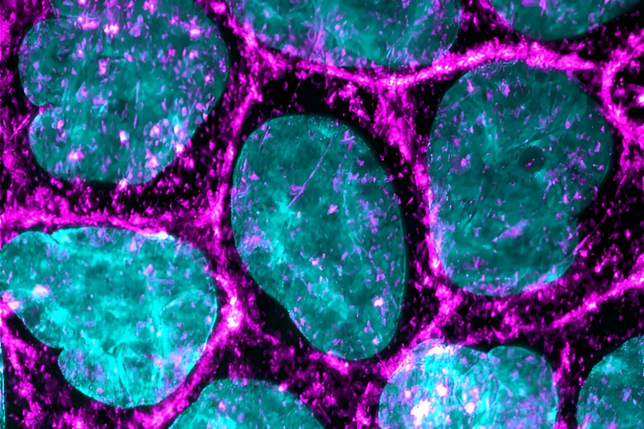
Microscopic image of the epithelial cells used in the study, the cytoskeleton (magenta) and nuclei (cyan) of the cell are fluorescently labeled, allowing detailed study of the structures. Credit: Heidi Peussa
Life sciences and photonics researchers at the University of Tampere have made a remarkable discovery by studying the response of surface cells to mechanical stimuli. By simulating the deformation of the extracellular matrix beneath cells, the researchers showed that cells quickly detect even minor changes in their environment and that their response is more complex than expected. This discovery could help to better understand, for example, the processes linked to the formation of cancer metastases.
As part of this joint project, three research groups studied how epithelial cells detect small changes in their environment via ion channels. The study was carried out using light-sensitive materials developed by the Smart Photonics Materials research group led by Professor Arri Priimägi, which can be used as a substrate for cell culture. These materials enable precise and controllable movement of the cellular substrate through light stimulation.
“The cells had a marker protein for intracellular calcium, so we were able to draw small grooves on the surface of the substrate on a confocal microscope and at the same time monitor how living cells react to these changes in the environment with the help calcium.” says Teemu Ihalainen, senior researcher at the Tampere Institute for Advanced Studies (IAS) and head of the Cellular Biophysics Research Group at the Faculty of Medicine and Health Technology.
“We found that the movement of even a few tens of nanometers of material opened mechanically closed calcium channels in cells, through which cells could change their calcium levels.”
Calcium is needed by cells for a wide variety of processes, so even small changes in the amount of calcium can have large effects on cellular functions. The study shows, perhaps for the first time, that cells are able to detect minute movements in their environment and that these movements are detected by changing the flow of calcium ions across the cell membrane, i.e. that is, electrically via ionic currents.
The study focused on intracellular changes in calcium during the first few seconds of the mechanical stimulus. A paper titled “Light-induced nanoscale deformation in a thin azobenzene film triggers rapid intracellular Ca.”2+ augmentation via mechanosensitive cation channels”, which constitutes a key element of the thesis of doctoral student Heidi Peussa, was published in the journal Advanced science.
Mechanically gated ion channel as key
In the body, epithelial cells are closely linked to the extracellular matrix, which makes it possible, for example, to transmit mechanical stresses from the environment to the cells. Mechanical stimuli are important in the normal functioning of cells. Disruption of cell attachment often causes disease or other problems.
Cells detect changes in their environment in a variety of ways, for example through mechanically gated PIEZO1 ion channels. Channels can be understood as pores in the cell membrane that are closed in a mechanically relaxed state, but open when the cell membrane stretches. The opening occurs in thousandths of a second and results in an influx of calcium into the cell. The process plays a key role in many physiological functions, for example in tactile sensation. The discovery of mechanically gated ion channels was awarded the Nobel Prize in 2021.
The study showed that PIEZO1 channels are essential for sensing rapid changes in the cellular microenvironment.
“We found that cells are able to detect deformations as small as 40 nanometers (0.000040 mm) that occur in thousandths of a second. For the first time, we were able to monitor how the PIEZO1 channel opens as a result of a physical change in the local extracellular environment,” explains Ihalainen.
In the study, calcium signals from cells cultured on DR1 photosensitive glass were monitored under the microscope using green light and, at the same time, mechanical stimuli with blue light were induced on the basal side of the cells. cells. Illustration: Heidi Peussa. Credit: Heidi Peussa, University of Tampere
New possibilities for studying cellular processes in the eye
The method used by the researchers is new and makes it possible to study in particular the mechanical stimulus of the extracellular matrix and to simultaneously monitor cellular responses. Further research into how the PIEZO01 channel works is already underway. Furthermore, the researchers’ goal is to study and develop new light-sensitive materials.
“Our next steps are to study the regulation and regulatory factors of these mechanically triggered ion channels. The goal is also to better understand what happens after the first few seconds in the perception of the sensation of force,” explains Soile Nymark , Associate Professor. of Biosensor Technology and Head of the Eye Biophysics Research Group at the Faculty of Medicine and Health Technology.
“We are developing new transgenic cell lines to study calcium signaling at different locations within the cell in more detail. These transgenic cell lines also allow us to extend studies to the underlying retinal pigment epithelium of the eye and d “study the role of PIEZO1 channels in the maintenance of the retina. “
More information:
Heidi Peussa et al, Light-induced nanoscale deformation in thin azobenzene film triggers rapid intracellular Ca2+ Increase via mechanosensitive cation channels, Advanced science (2023). DOI: 10.1002/advs.202206190
Provided by the University of Tampere
Quote: Study shows that cells respond quickly to small movements in the microenvironment induced by light (January 25, 2024) retrieved January 25, 2024 from
This document is subject to copyright. Apart from fair use for private study or research purposes, no part may be reproduced without written permission. The content is provided for information only.
