

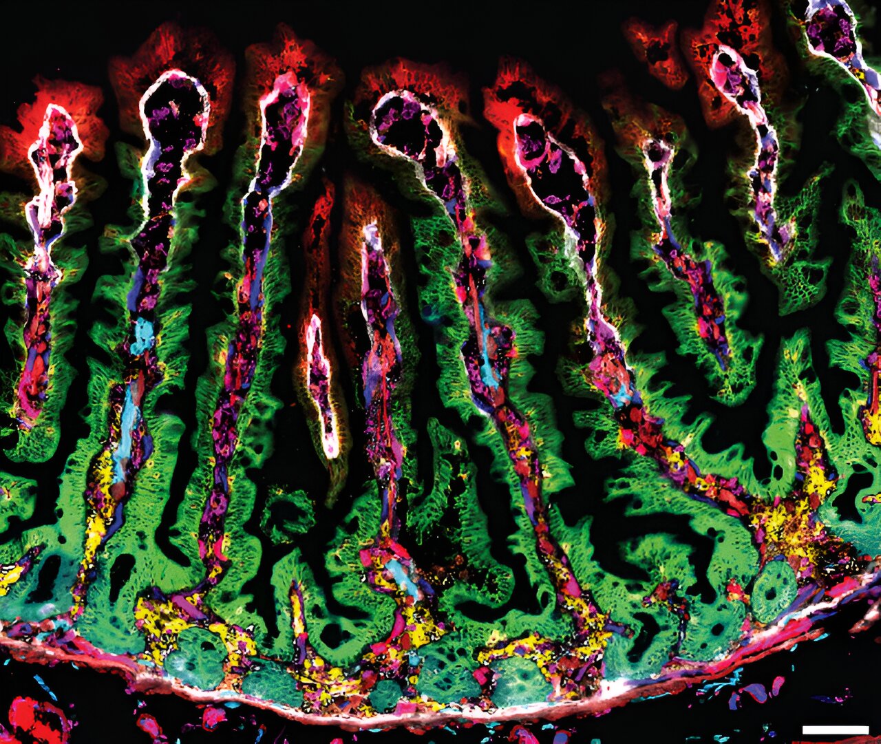
Image of human small intestinal tissue obtained using fluorescent dyes that labeled many proteins in parallel, mapping the different proteins produced in each part of the intestinal villi. Credit: Weizmann Institute of Science
Tourists visiting an unfamiliar city would have difficulty finding their way around using only a topological map, no matter how detailed it may be. Most tourist maps therefore highlight interesting sites and important monuments.
Already in the 16th century, anatomy books described the obscure territory of the human small intestine. We know, for example, that this digestive tract is on average 6 meters long and is covered with millions of villi, tiny finger-like projections that multiply the surface area of the tube thirty-fold and are separated by crypt-like crevices.
Until now, it was unclear exactly where the interesting sites and important landmarks were located in this complex configuration of crypts and villi. In a new study, reported in NatureResearchers from the Weizmann Institute of Science and experts from the Department of General Surgery at Sheba Medical Center have compiled the first detailed map of the different activity zones of the human small intestine, revealing what makes it so efficient at absorbing nutrients and protecting the body from infections.
Conditions along the small intestine vary dramatically from the crypts lining the inner walls to the tips of the protruding villi. While the area closest to the small intestine wall has an abundant supply of blood and oxygen, the environment at the tips of the villi is low in oxygen and saturated with nutrients and bacteria.
In 2018, a research team led by Professor Shalev Itzkovitz of Weizmann’s Department of Molecular Cell Biology showed that cells in the villi of the small intestine of mice adapt to a changing environment and perform defined functions depending on their relative location on the villi.
“Until then, we had only worked with mice,” says Dr. Yotam Harnik, who completed his PhD in Itzkovitz’s lab, recounting the origins of the project to build a genetic atlas of the human small intestine.
“One day I was having lunch on the lawn of the Weizmann campus and talking with Dr. Oran Yakubovsky, a Sheba surgical resident who had started his PhD in our lab six months earlier. He asked me why we didn’t harvest human intestinal tissue in the operating room. One of the problems with this practice is that surgeons often don’t harvest a significant portion of a person’s intestine when they’re healthy.”
“We decided to analyze human intestinal tissue, using samples from Whipple procedures, in which the head of the pancreas is removed due to pancreatic pathologies,” Yakubovsky explains.
“In this procedure, surgeons also remove the entire duodenum, the first part of the small intestine. One of the advantages of this procedure from a research perspective is that the intestinal tissue, which is removed for anatomical reasons, is considered healthy and can be used to study the normal intestine. We were able to cooperate with the Sheba General Surgery Department, ensuring that each sample, in its entirety, was frozen quickly.”
At the same time, the Weizmann Institute has acquired new technology that allows researchers to efficiently map spatial gene expression in tissues and analyze, with a resolution of 50 microns, which genes are expressed in each region and to what extent.
Slow release fats
The atlas that the researchers compiled, led by Harnik and Yakubovsky, both of Itzkovitz’s lab, in cooperation with Dr. Rouven Hoefflin of Professor Itay Tirosh’s lab in Weizmann’s Department of Molecular Cell Biology, sheds light on some of the long-standing mysteries surrounding how the small intestine works.
Already in the 1950s, scientists had discovered that it takes up to two days for dietary fats to be absorbed into the bloodstream, which prevents an increase in blood fat levels. However, the exact mechanism of this phenomenon was not yet well understood. The atlas of the human intestine shows that the digestion of fats by the human villi resembles an assembly line.
Cells deep in the villi lock up the dietary fat in fat droplets, and it is only several hours later, when these cells move along the villi and reach the tip, that they load the fat onto “cargo trucks,” huge particles that carry it through the lymphatic system to the blood vessels, and from there to storage in the body.
The regulation of iron balance in our body is also like an assembly line: iron is absorbed into the crypts and deep into the villi; when the cells reach the tip of the villi (and depending on the iron level in the body), they release their iron reservoir into the blood or take it with them to their death in the intestinal cavity.
The atlas also reveals that absorption and production of enzymes needed to digest other important nutrients (amino acids, short proteins, and sugars) occurs only at the tips of the villi, while cells at the base of these protuberances specialize in the absorption of vitamins and minerals.
As for the immune defense provided by the small intestine, the researchers discovered that cells located at the tips of the villi secrete antimicrobial proteins that directly attack bacteria and also send a call for help to the most aggressive cells of the immune system. The tips of human villi were thus found to be rich in immune cells that promote inflammation.
Epithelial gene expression programs zoned along the crypt-villus axis. Credit: Nature (2024). DOI: 10.1038/s41586-024-07793-3
The long and winding route
Anatomy textbooks traditionally describe the villi of the healthy human small intestine as straight, finger-like protrusions. However, when compiling their atlas, the researchers identified villi that branch off from other villi, something that had previously only been observed in cancerous tumors. The scientists believe that this branching serves to further increase the surface area of the small intestine and improve absorption. The researchers achieved these results using a new method that allows researchers to document the spatial structure of tissues in three dimensions without disrupting the integrity of the tissue.
“Our atlas provides answers to some fundamental research questions, but it can also be applied to clinical questions,” Itzkovitz says. “Now that we have mapped a healthy small intestine, we can begin to better understand the changes it undergoes when it is diseased, when we age, when we take certain medications, or when we follow a specific diet.”
“This study, like others we are currently conducting in the Sheba Department of General Surgery, is only possible because the senior members of the department, led by Dr. Ido Nachmany, appreciate the importance of basic research. This framework allows Weizmann physician-scientists to translate questions that arise in the clinic into studies that can provide answers.”
Nachmany, Director of the Sheba Department of General Surgery, adds: “The cooperation between the Weizmann Institute and the Sheba Department of General Surgery has resulted in a series of fascinating studies on the most fundamental aspects of the physiological and anatomical functioning of human tissues.
“The collaboration between the two worlds – basic research and clinical medicine – is a tremendous achievement, and it is reflected in this study NatureWe are working to strengthen this connection and hope that more surgical residents will devote significant time to research at the institute. We are convinced that this will have considerable benefits for both worlds.
More information:
Yotam Harnik et al., A spatial expression atlas of the human adult proximal small intestine, Nature (2024). DOI: 10.1038/s41586-024-07793-3
Provided by the Weizmann Institute of Science
Quote:Navigating the Digestive Tract: Study Proposes First Detailed Map of the Small Intestine (2024, August 29) Retrieved August 29, 2024 from
This document is subject to copyright. Apart from any fair dealing for the purpose of private study or research, no part may be reproduced without written permission. The content is provided for informational purposes only.
