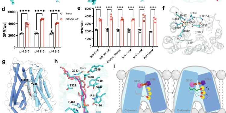
Structural basis of S1P transport via human SPNS2. A Overall structures of SPNS2 in apo, S1P-bound, FTY720-P-bound, and 16d-bound states. Left panel, orthogonal views of the cryo-EM density map; right panel, a model of the complex in the same view. b The hydrophobic pocket for the lipophilic tail of S1P and FTY720-P. The yellow surface color indicates a hydrophobic region and the blue color indicates a hydrophilic region. vs The detail of the hydrophobic pocket for fixing the substrate. d The effect of extracellular pH change on SPNS2 transport activity. e The effect of changing the extracellular concentration of potassium and sodium on the transport activity of SPNS2. F A polar interaction network on the extracellular side of SPNS2. g A gate formed by the hydrogen bonding interaction between Y246 and G333 to lock SPNS2 in an inward-facing state. h A hydrophilic tunnel filled with polar residues under gate G333/Y246. I A proposed “ladder” mechanism for S1P transport via SPNS2. j Transport activities of SPNS2 mutants. Data are presented as mean ± SD; not= 3 independent samples; ns, no significance; *P. <0.05; ** P. <0.01; * **P.<0.001,****P.<0.0001. One-way and two-way ANOVAs were used. Credit: Cellular research(2023). DOI: 10.1038/s41422-023-00913-0
When an enemy invades, defenders are transported to neutralize the marauders. In the human body, a protein transporter called SPNS2 carries S1P molecules from endothelial cells to rally the immune cell response in infected organs and tissues.
Using specially developed nanobodies that bind to SPNS2 and enlarge the entire structure, the enlarged SPNS2 structure allows visualization of S1P molecules by cryogenic electron microscopy. Scientists from the Translational Immunology Research Program at the National University of Singapore’s Yong Loo Lin School of Medicine and their partners have analyzed the structure of the SPNS2 protein at the atomic level, which could provide better insights into how S1P signaling molecules are released to communicate with immune cells to regulate inflammatory responses.
“Seeing is believing. This work shows that SPNS2 directly exports S1P for signaling and that it is possible to inhibit its transport function with small molecules. This work provides the basis for understanding how S1P is released by SPNS2 and how this protein function is inhibited by small molecules for the treatment of inflammatory diseases,” said team leader Dr. Nguyen Nam Long.
The SPNS2 protein allows the binding of S1P signaling molecules to trigger immune cells to leave lymph nodes and induce inflammation in different parts of the body when needed. Made up of amino acids, the SPNS2 protein is malleable enough to change shape and structure to release S1P signaling molecules through small cavities within the protein.
With the discovery of how the SPNS2 protein releases S1P molecules, the SPNS2 structure can be exploited for future drug development. Similar to discovering the shape of the lock before the key was designed, this discovery sheds more light on how future drugs can be designed to better target the protein to increase drug effectiveness.
This finding builds on previous research, which showed that removing the SPNS2 protein from a preclinical model effectively blocks the S1P signaling pathway, so that S1P signaling molecules cannot be transported to induce cells immune cells to leave the lymph node to induce inflammation. The SPNS2 protein and the signaling molecule S1P are required for the recruitment of immune cells to inflammatory organs, which serves the treatment of various inflammatory diseases.
“Using preclinical models, we showed that targeting SPNS2 proteins in the body blocks inflammatory responses in diseases such as multiple sclerosis. This work gave us the opportunity to inhibit its transport function with small molecules that will go a long way to treat inflammatory diseases more effectively and efficiently,” said Dr. Nguyen.
The document was published in Cellular researchin December 2023.
More information:
Yaning Duan et al, Structural basis of sphingosine-1-phosphate transport via human SPNS2, Cellular research(2023). DOI: 10.1038/s41422-023-00913-0
Provided by the National University of Singapore
Quote: SPNS2 directly exports S1P for signaling, can be inhibited (February 15, 2024) retrieved February 15, 2024 from
This document is subject to copyright. Apart from fair use for private study or research purposes, no part may be reproduced without written permission. The content is provided for information only.


