

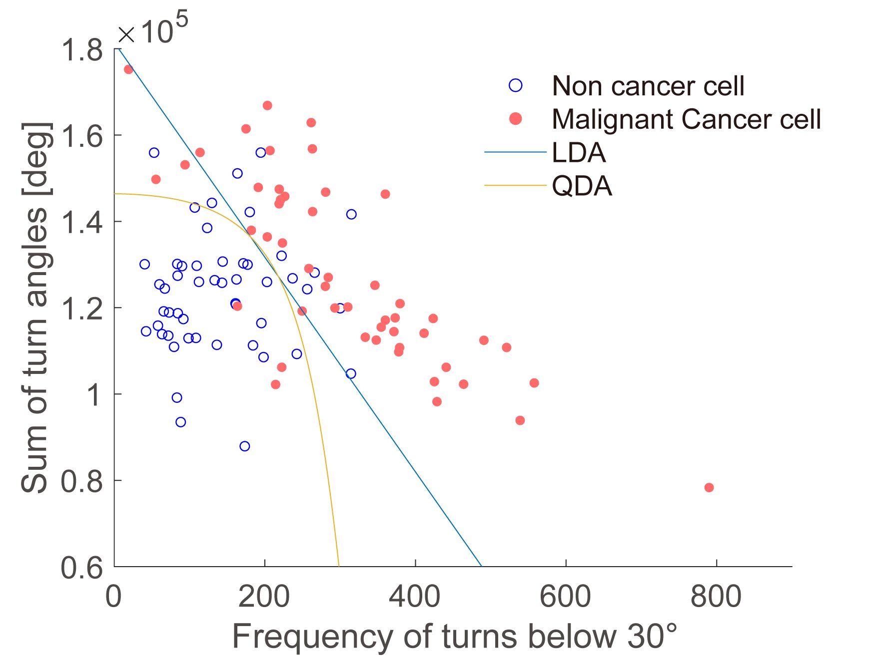
Discriminate the state of the cell using the “curvature” of the trajectories. The sum of the cornering angles and the frequency of shallow turns can be used to separate healthy and cancerous cells. The lines show different ways to identify the border between the two cell classes. Credit: Tokyo Metropolitan University
Researchers from the Tokyo Metropolitan University have discovered that the movement of unmarked cells can be used to say whether they are cancer or healthy. They observed malignant fibrosarcoma cells and healthy fibroblasts on a dish and have found that monitoring and analyzing their paths can be used to differentiate them with up to 94% precision.
Beyond the diagnosis, their technique can also shed light on the functions related to cell motility, such as the healing of the tissues. The document is published in the journal Plos a.
While scientists and medical experts have examined cells under the microscope for many centuries, most studies and diagnoses focus on their form, which they contain and where different parts are inside. But the cells are dynamic, change over time and are known to be able to move.
By following and analyzing their movement with precision, we can be able to differentiate cells which have functions based on cell migration. An important example is the metastases of cancer, where the motility of cancer cells allows them to spread.
However, this is easier to say than to do. On the one hand, the study of a small subset of cells can give biased results. Any precise diagnostic technique would be based on automated and high speed monitoring of a significant number of cells.
Many methods then turn to fluorescent marking, which makes cells much easier to see under the microscope. But this labeling procedure can itself affect their properties. The ultimate objective is an automated method that uses conventional microscopy without an label to characterize the motility of the cells and show whether the cells are healthy or not.
Draw the paths of cancer and healthy cells. The team used phase contrast microscopy to identify the trajectories of individual cells. Credit: Tokyo Metropolitan University
Now, a team of researchers from Tokyo Metropolitan University led by Professor Hiromi Miyoshi has found a way to follow cells using phase contrast microscopy, one of the most common ways to observe cells.
Phase contrast microscopy is entirely without label, allowing cells to move on a petri box closer to their natal state, and is not affected by the optical properties of plastic kneading boxes through which the cells are colorful.
Thanks to an innovative image analysis, they were able to extract the trajectories from many individual cells. They focused on the properties of the paths taken, such as the speed of migration, and how the paths were curved, which would all code subtle differences in deformation and movement.
As a test, they compared the healthy fibroblast cells, the key component of the animal tissue and the cells of malignant fibrosarcoma, the cancer cells which derive from the fibrous connective tissue. They were able to show that the cells have migrated subtly differently, as characterized by the “sum of the corners of turns” (how winding the paths), the frequency of shallow turns and the speed with which they moved.
In fact, by combining both the sum of the corners of turn and the frequency they made shallow turns, they could predict whether a cell was cancerous or not with a precision of 94%.
The team’s work promises not only a new way of discriminating cancer cells, but also applications in search of any biological function based on cell motility, such as healing of injuries and tissue growth.
More information:
Sota Endo et al, development of cells monitoring without label for the discrimination of heterogeneous mesenchymal migration, Plos a (2025). DOI: 10.1371 / Journal.pone.0320287
Supplied by Tokyo Metropolitan University
Quote: Scientists can distinguish healthy and cancer cells by the way they move (2025, April 21) recovered on April 21, 2025
This document is subject to copyright. In addition to any fair program for private or research purposes, no part can be reproduced without written authorization. The content is provided only for information purposes.
