

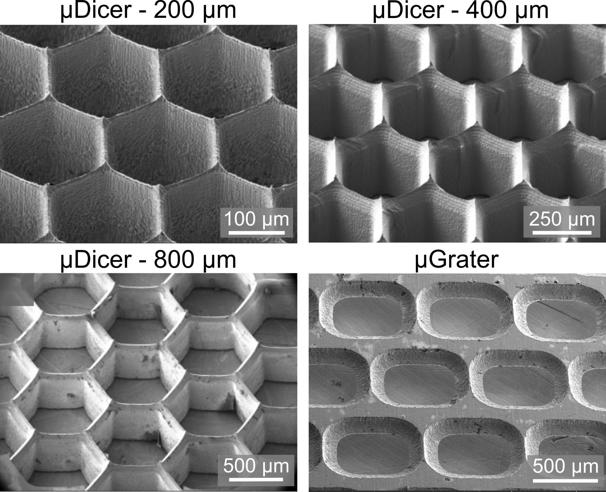
Scanning microscopy images of the microDicer (blade spacings of 200 μm, 400 μm, and 800 μm, respectively) and microGrater. Credit: Seth Cordts and Saisneha Koppaka
As fascinating as working in a modern biology lab is, it often requires repetitive, detailed work before research can begin. For example, cancer researchers are now able to use hundreds or even thousands of small, lab-grown tumor samples, called organoids, to test multiple cancer therapies, including immunotherapies, at once.
To produce organoids, researchers often have to hand-cut a fresh tumor into small pieces, using scissors to slice, slice, slice the specimen down to sub-millimeter size. This dissection work is tedious and yet often done by graduate students or trained, and usually overqualified, researchers.
That tedious period may soon be over, as researchers at the Stanford School of Engineering have developed two new tools that speed up the precise cutting of tumor samples into organoids at the submillimeter scale. Introducing the microDicer (µDicer) and microGrater (µGrater).
The inventors believe that these tools, which are reminiscent of familiar utensils used in kitchens around the world to dice vegetables or grate cheese, will allow researchers to improve sample consistency and quality, which directly impacts the quality of data from downstream experiments, such as drug response measurements.
“Our collaborator and co-author Calvin Kuo, co-director of the Cancer Biology Program at Stanford Cancer Center, has shown that in cancer immunotherapy research, maintaining the spatial relationships between groups of tumor cells and immune cells that have already infiltrated the tumor, as opposed to working with individual cells, is really important for the accuracy of drug and immunotherapy testing. Our tools help researchers more efficiently create organoids that maintain these relationships,” says Sindy Tang, associate professor of mechanical engineering and senior author of a new study describing the microDicer and microGrater.
The work appearsMicrosystems and nanoengineering.
“These new tools will speed up manual work in the lab, but their usefulness goes beyond this obvious advantage,” Tang added. “These tools produce organoids of uniform size, and the slides can be tailored to the size the researcher wants.”
Precise control of organoid size allows researchers to ask whether there is a “perfect” organoid size. If the organoid is too small, it may not retain the original properties of the tumor as it existed in the body. However, if it is too large, it may die because oxygen and nutrients have difficulty reaching the tumor cells inside.
Presentation of the µDicer and µGrater devices and first tests with 2% agarose and porcine kidney tissue. Credit: Microsystems and nanoengineering (2024). DOI: 10.1038/s41378-024-00756-8
Precise blades
Tang and his team fashioned the microDicer blades out of silicon using conventional micromachining tools used in computer chip manufacturing. The blade patterns are etched into the silicon using a reactive plasma. The microGrater blades are made of stainless steel.
The microDicer blade resembles a mesh of nested vertical hexagons, resembling a honeycomb with sharp edges. To use the microDicer, a researcher cuts thin layers of tissue that are then pressed through the microDicer’s honeycomb mesh to produce precise, uniform tumor samples.
The microGrater’s blade array, meanwhile, features a series of sharp, rounded rectangles just over half a millimeter long; the edges of each rounded rectangle are beveled to form the blades that shave precise organoids as the tissue is moved back and forth across the rasp.
Promise of the future
The tumor samples Tang and his colleagues studied are grown in lab mice and are a good model for human tumors. The ultimate goal is a kind of personalized, tailored cancer therapy in which samples are taken from a specific patient to test which immunotherapies work best for them. By standardizing the process flow and organoid sizes in a way that manual chopping doesn’t, these new tools could speed approval by regulators, like the FDA, for broader use, Tang says.
Her most recent work, which leverages these technologies, aims to identify the ideal size of organoids for different tissue types. It is possible that different cancers and tissues have different dimensional requirements, something that the flexibility of Tang’s tools should help address. She can adjust the blades to make organoids of virtually any size, from a few hundred micrometers to a millimeter. In addition, she is currently studying how the shape of the blades affects the fidelity of the sections. The physics of cutting soft biological tissues is a difficult and poorly understood problem.
Once Tang had the idea for the microDicer, she turned the prototyping process into a graduate-level product design course, called the Advanced Micro and Nano Fabrication Laboratory, taught by Roger Howe and Jonathan Fan. In the course, PhD students learned how to fabricate microscale devices in a hands-on, project-based environment at the Stanford Nanofabrication Facility. Several of Tang’s students are co-authors on the paper.
The microDicer and microGrater aren’t Tang’s first mechanical tools for biological purposes. She made headlines a few years ago with another tool, a cell guillotine, which could split individual cells in half to study how individual cells heal. That invention sparked considerable interest from other biology labs interested in the possibility of cutting tissue, which prompted the development of the microDicer and microGrater. She is currently evaluating ways to disseminate these tools within the scientific community, including the possibility of commercializing them.
More information:
Seth C. Cordts et al, Microdissection tools for generating organoids to model the tumor immune microenvironment, Microsystems and nanoengineering (2024). DOI: 10.1038/s41378-024-00756-8
Provided by Stanford University
Quote:New precision tools enable rapid tumor dissection (2024, September 10) retrieved September 10, 2024 from
This document is subject to copyright. Apart from any fair dealing for the purpose of private study or research, no part may be reproduced without written permission. The content is provided for informational purposes only.
