

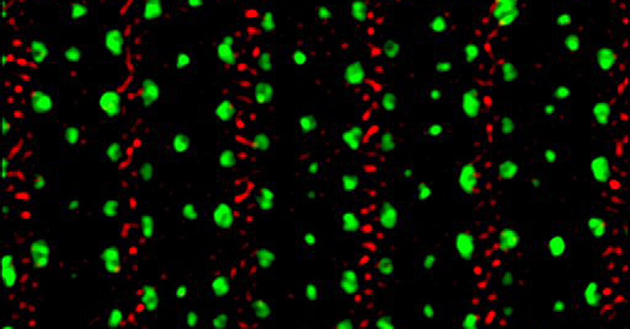
Super-resolution microscopy reveals two roundworm collagens marked in red and green. Credit: Natural communications (2023). DOI: 10.1038/s41467-023-43058-9
Species in the animal kingdom present vital interfaces between the outermost layers of their bodies and the environment. Complex microscopic structures – found on the outer layers of human skin, for example – are known to assemble in matrix patterns.
But how these complex structures, known as apical extracellular matrices (aECMs), are assembled into elaborately woven architectures remains an elusive question.
Now, after years of research and the power of a technologically advanced instrument, scientists at the University of California, San Diego have discovered the basis of these matrices in a tiny nematode. The roundworm Caenorhabditis elegans has been the subject of much study for decades because of its transparent structure that allows researchers to look inside its body and examine its skin.
Described in the newspaper Natural communications, researchers from the School of Biological Sciences have now deciphered the assembly of aECM patterns in roundworms at the nanoscale. A powerful super-resolution microscope revealed novel patterns related to the columns, called struts, which are essential for the proper development and function of aECMs.
“The struts are like tiny pillars that connect the different layers of the matrix and serve as scaffolding,” said Andrew Chisholm, professor in the Faculty of Biological Sciences and lead author of the paper.
A 3D image reveals struts (green) alongside collagens (magenta). Credit: Natural communications (2023). DOI: 10.1038/s41467-023-43058-9
Although roundworms serve as a model organism for laboratory studies due to their simple, transparent bodies, beneath the surface they exhibit complex architectures. They also have nearly 20,000 genes, a number comparable to the number of human genes, and therefore provide insights into the structure and function of more advanced organisms.
Focusing on the roundworm exoskeleton known as the cuticle, the researchers found that defects in the struts led to abnormal swelling of the layer, or “blisters.” Within the cuticular layer, the study focused on collagens, which are the most abundant family of proteins in our body and help hold body materials together.
“Struts hold critical layers together,” Chisholm said. “Without them, the layers separate and cause disorders such as blisters. In blistered mutants, you don’t see any spacers.”
Conventional laboratory instruments had previously photographed the struts without detail, often resulting in undefined spots. But thanks to the laboratory of Assistant Professor of Biological Sciences Andreas Ernst, they gained access to advanced instrumentation, known as 3D Structured Illumination Super-Resolution Microscopy (3D-SIM), which brought the struts into focus and enabled to define their functions more easily. The researchers were then able to resolve the nanoscale organization of the struts and previously undocumented levels of patterning in the cuticular layer.
“We could see exactly where these proteins were going in the matrix,” Chisholm said. “It’s potentially a paradigm for how the matrix assembles into very complex structures and very complex patterns.”
The first two authors, Jennifer Adams (senior research associate) and Murugesan Pooranachithra (postdoctoral fellow), contributed equally to the article. Other co-authors are Erin Jyo, Sherry Li Zheng, Alexandr Goncharov, Jennifer Crew, James Kramer, Professor of Neurobiology Yishi Jin, Assistant Professor of Cellular and Developmental Biology Andreas Ernst and Andrew Chisholm.
More information:
Jennifer RG Adams et al, Nanoscale modeling of collagens in the apical extracellular matrix of C. elegans, Natural communications (2023). DOI: 10.1038/s41467-023-43058-9
Provided by University of California – San Diego
Quote: Inside the Matrix: Nanoscale Patterns Revealed Within a Model Research Organism (December 21, 2023) retrieved December 21, 2023 from
This document is subject to copyright. Apart from fair use for private study or research purposes, no part may be reproduced without written permission. The content is provided for information only.
