Credit: Current biology (2024). DOI: 10.1016/j.cub.2024.01.016
Cornell neuroscientists have identified a group of midbrain neurons that are essential for the social vocalizations mice produce, but not for the squeaks they make when in distress.
The results suggest that in mice, and likely in other animals, different populations of midbrain neurons control different types of vocalizations, rather than a single multitasking set.
A better understanding of how the brain is organized to control vocalization, including human speech, could reveal how genetic disorders or diseases can damage these neural circuits and improve treatments.
“Vocal communication is central to our experience as human beings and fundamental to the social success of animals in general,” said Katherine Tschida, Mary Armstrong Meduski ’80 assistant professor in the Department of Psychology in the College of Arts and Sciences. science. “We are beginning to uncover in detail how different populations of neurons contribute to specific aspects of our vocal behaviors.”
Tschida is the corresponding author of “Midbrain neurons important for producing mouse ultrasonic vocalizations are not required for distress calls,” published Jan. 31 in Current biology. First author Patryk Ziobro, a doctoral student in the field of psychology, and co-authors Yena Woo and Zichen He are current and former members of the Tschida Lab.
Scientists have known for decades that a part of the midbrain called the periaqueductal gray (PAG) is essential for vocal production, which can include a cat’s meow or hiss, a person’s laugh or cry, or the content emotional of our speech. This knowledge remained at the regional level, Tschida said, without the ability to selectively manipulate the neurons responsible for vocalization.
Mice communicate primarily in two ways: human-audible squeaks when they feel pain or fear; and high-frequency ultrasonic vocalizations (USV) used during courtship and other social interactions. In 2019, a team led by Tschida reported that destroying a particular group of PAG neurons cut off males’ ultrasonic communication, raising the question of whether these neurons could control vocalizations in other contexts.
The new research replicated the previous study’s findings in men and extended them to women. Lacking what the researchers called PAG-USV neurons, the mice could still engage in social and courtship activities, but did so silently.
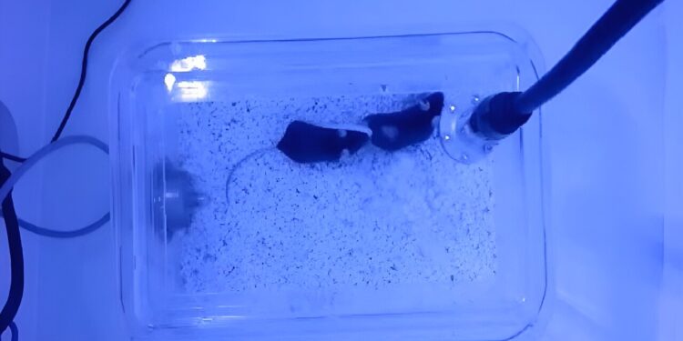
The team then looked to see if the absence of neurons also limited the mice’s ability to squeak. By applying mild shocks to the study subjects’ feet, enough to cause squeaking but cause no physical harm, the researchers found that distress calls were unaffected.
“The fact that you can eliminate one type of vocalization without disrupting the other is pretty clear evidence that there must be different neurons in the brain regulating these two types of vocal communication,” Tschida said.
Ablation of PAG-USV neurons blocks USV production in male and female mice. (A) Schematic shows the experimental timeline for TRAP2-mediated ablation of PAG-USV neurons and behavioral measurements. (B) Total USVs produced by experimental (PAG-USVcasp, black dots, n = 11) and control (PAG-USVGFP, green dots, n = 12) male mice during 30 min social interactions with females are shown before and after 4-OHT treatment. Significant main effects are indicated by text above the graph, and significant interaction effects are indicated in the graph. (C) Same as (B), for the proportion of time men spent interacting with women before and after 4-OHT treatment. (D) Same as (B), for the number of USVs produced per second of male-initiated social interaction before and after 4-OHT treatment. (E) Total USVs produced by experimental (PAG-USVcasp, black dots, n = 12) and control (PAG-USVGFP, green dots, n = 12) female mice during social interactions with females are shown before and after 4-OHT treatment. . (F) Same as (E), for the proportion of time women spent interacting with women before and after 4-OHT treatment. (G) Total USVs are plotted for interactions between pairs of females including 1 PAG-USVGFP female and one intact female (left, n = 12 trials), 1 PAG-USVcasp female and one intact female (middle, n = 12 trials) and 2 PAG-USVcasp females (right, n = 8 trials, including round-robin pairings of n = 6 PAG-USVcasp females). The data in the left and middle columns are the same data represented in (E), post-4-OHT. Credit: Current biology (2024). DOI: 10.1016/j.cub.2024.01.016
The scenario might be more complex in animals with richer vocal repertoires, Tschida said, including humans. But the research seems to rule out simple models suggesting that a single population of neurons is causing it all.
It’s a step, Tschida said, toward building a more detailed mechanistic picture that explains how PAG neurons work specifically and how interactions with downstream neurons in the hindbrain allow animals to vocalize at the right time and to the right way. And, researchers say, it’s a step toward understanding why neurological and neurodevelopmental disorders can impair vocalization.
“In cases where this impairment is more related to motor control, we do not clearly understand what causes the difficulties developing and regulating speech,” Tschida said. “Having this basic level of understanding about how the brain generates vocalizations can help us understand when something is wrong, how did it happen?”
Interestingly, Tschida said, the study also showed that different neurons are responsible for vocalizations conveying positive and negative emotional states in mice, which could be another avenue of research. In ongoing work, the team hopes to identify the neurons that regulate the production of squeaks.
“Knowing that there are different populations of neurons for USVs and squeaks opens the way to a whole new set of experiments delving into the neural mechanisms of these vocalizations,” Ziobro said. “I look forward to the next steps.”
More information:
Patryk Ziobro et al, Midbrain neurons important for producing mouse ultrasonic vocalizations are not necessary for distress calls, Current biology (2024). DOI: 10.1016/j.cub.2024.01.016
Provided by Cornell University
Quote: Mouse social calls and distress calls are linked to different neurons, new research shows (January 31, 2024) retrieved January 31, 2024 from
This document is subject to copyright. Apart from fair use for private study or research purposes, no part may be reproduced without written permission. The content is provided for information only.


