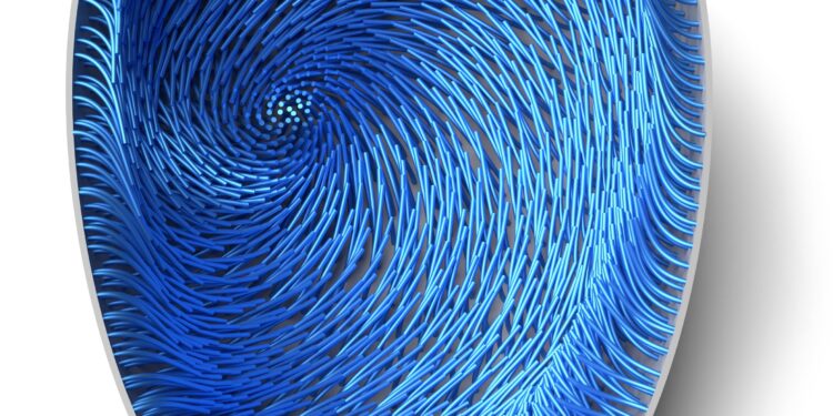
Simulation carried out at the Center for Computational Biology (Flatiron Institute) using their SkellySim simulation software. Credit: Dutta et al.
Cytoplasmic flow is the large-scale movement of cytoplasm (i.e., gelatinous fluid inside cells) within a living cell. This flow, known to regulate various intracellular processes, can vary significantly between different cell types at different stages of cellular development. Examining and modeling different types of cytoplasmic flows can help us understand how they emerge in specific cell types.
Previous studies have primarily examined continuous cytoplasmic flows in large cells where, as is often claimed, diffusion is too slow to enable the biological processes necessary for organisms to perform (e.g., the development of an egg or of an embryo, or in large plant cells).
Thanks to this slow diffusion, the flow allows faster distribution of cellular components. In early fly oocytes (i.e. developing ovules), for example, cytoplasmic flow appears random, while at later stages of development, where the oocyte is larger, they may appear to large scale and rotating.
Researchers at the Flatiron Institute, building on previous work, recently introduced a versatile modeling strategy that can be used to study self-organized cytoplasmic flow in systems consisting of hydrodynamically coupled deformable fibers.
This model, introduced in Natural physics and in collaboration with scientists from Princeton and Northwestern Universities, was combined with data collected in experiments on the Drosophila (i.e. fruit fly) oocyte to gather information on the self-organized cytoplasmic flow.
“I have been working for some time in the general areas of biologically active matter, intracellular mechanics, and complex fluids,” Michael J. Shelley, co-author of the paper, told Phys.org. “The problem addressed in our recent paper combines all of these areas, each of which appeals to me greatly.
“I learned about this particular problem of flows in oocytes from my friend Ray Goldstein and realized that previous work with my colleague at Flatiron, David Stein, could be adapted to understand something about the problem of oocytes. This is what happened, and David and I worked with Ray and his colleagues at Cambridge on a very refined first 2D model.” This work was published in Physical Examination Letters in 2021.
Flatiron Institute researchers previously developed various tools to study the hydrodynamics of moving microtubules, rigid biopolymers that constitute a central part of the cellular cytoskeleton. Shelley, Stein and their colleagues Reza Farhadifar, Sayantan Dutta and Stas Shvartsman planned to use these digital tools to study the appearance of self-organized cytoplasmic flows in 3D cells.
“The main goal of our recent study was to provide a minimal, but not too minimal, model, invoking only microtubules, molecular motors and the cytoplasm, that could explain experimental observations and help make predictions,” explained Shelley .
The recent study by Shelley and colleagues combines physical and mathematical theories with experimental results. The researchers began by creating a model that they could then use to simulate self-organized cytoplasmic flow in the Drosophila oocyte.
“We wrote a mathematical model for the constraints that molecular motors create when moving on a microtubule,” Shelley said. “This model should allow the microtubule to bend under loads and, for its curvature, displace the cytoplasm, which affects the curvature of other microtubules. Next, we used high-quality software, here called SkellySim, which you allows you to simulate a few thousand of these microtubules interacting by collectively pushing fluid as they bend collectively.
After developing their model and running simulations, Shelley and his colleagues conducted experiments on Drosophila oocytes. First, they used optical microscopy to examine cytoplasmic movements in developing oocytes, then analyzed the data collected using particle imaging velocimetry to reconstruct the cytoplasmic velocity fields.
“Our paper provides a clear example of how, with very few ingredients, a large-scale transport system (i.e. continuous flow) could emerge in the cell from the interactions of a few components only (i.e. microtubules, motors, and cytoplasm),” Shelley said. “The beauty is in its robustness, because in much of the parameter space controlling the model, the system simply wants to form a tornado. This is, I think, a great example of biological self-organization to accomplish a stain.”
Notably, thanks to their model, the researchers were also able to predict the effect of cell shape on the orientation of tornadoes. Their predictions suggest that although the dynamics of cytoplasmic flow in Drosophila oocytes might be incredibly complex, they ultimately result in a simple end state (i.e., a tornado).
The findings gathered by Shelley and colleagues may soon pave the way for new explorations of cytoplasmic streaming, focusing specifically on this simple twisting state. This could lead to exciting new discoveries about the physics underlying life processes in biological cells.
“This work demonstrated the power that high-performance computing and modern algorithms can bring to understanding biophysical phenomena,” Shelley added. “In our next studies, we plan to explore how these flow twisters mix components throughout the cell or enable their delivery from one point to another.
“There are other transport systems within oocytes, such as ring channels, which are very interesting. I am generally interested in the multiple ways in which the cellular cytoskeleton is organized to accomplish cellular tasks.”
More information:
Savantan Dutta et al, Self-organized intracellular twisters, Natural physics (2024). DOI: 10.1038/s41567-023-02372-1
© 2024 Science X Network
Quote: Modeling and simulation of self-organized intracellular twisters in the Drosophila oocyte (February 21, 2024) retrieved February 21, 2024 from
This document is subject to copyright. Apart from fair use for private study or research purposes, no part may be reproduced without written permission. The content is provided for information only.


