

Credit: Nucleic acid search (2025). Doi: 10.1093 / NAR / GKAF152
Researchers from the Nano Life Science Institute (WPI-Nanolsi), the University of Kanazawa and colleagues carried out a breakthrough in understanding the DNA packaging of sperm. Using microscopy by high-speed atomic force (HS-AFM), they captured the process in real time of DNA condensation induced by protamin (PRM), providing critical information on fertility, stability of the genome and future medical applications. Their results are published in Nucleic acid search.
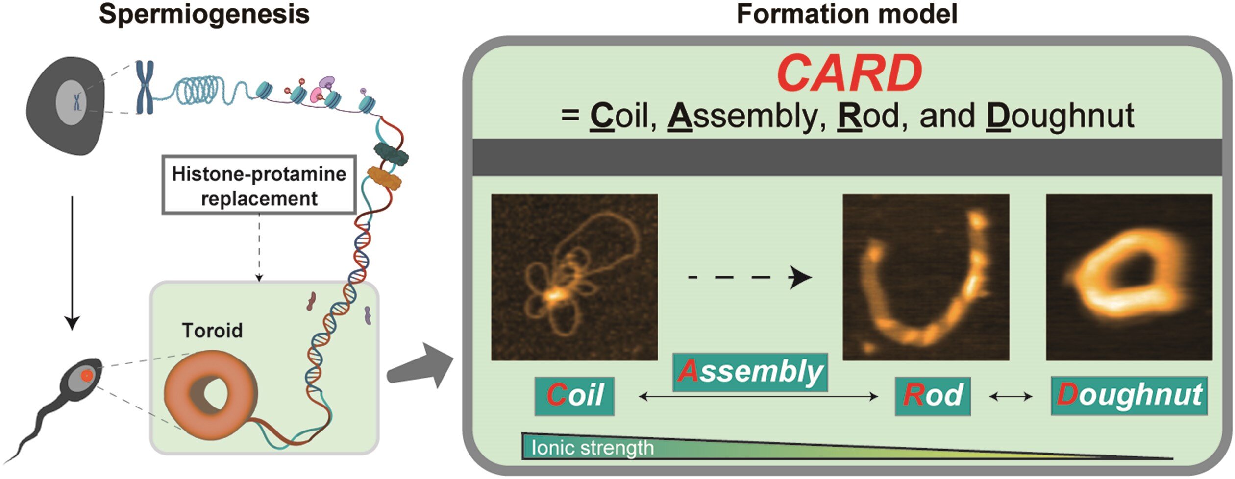
In most cells, DNA is rolled up around proteins called histones, which allows it to be vaguely packed and accessible for gene activity. However, in sperm, histones are replaced by protamins, which allows an extreme DNA condensation. This compaction is essential to protect genetic materials during fertilization, ensure effective DNA transport to egg and contribute to the development of fertility and embryo.
Despite its importance, the precise stages of the way in which the protamins condense DNA in highly stable structures have remained clear. Previous imaging methods could only capture static snapshots, leaving many unanswered questions. Now, for the first time, Richard W. Wong at the Nano Life Science Institute (WPI-Nanolsi), at the University of Kanazawa and collaborators used real-time imagery to reveal the entire condensation process.
Using HS-AFM, the search team directly viewed the stages transformation of DNA structures when they bind to protamins. The study introduces a new model of card card), which describes the condensation process through four distinct stages: the coil stage, where DNA forms loose loops; The assembly stage, where protamins bind, increasing the structural organization; The stem stage, where DNA becomes still compacted; And the Bignet stadium (Toroid), where the stable final structure forms.
In addition, researchers have discovered that this packaging is reversible, which means that the structure can move depending on environmental conditions. These ideas have major implications for understanding male infertility, chromatin biology and gene therapy.
Fertility research could benefit from information on DNA packaging, helping to diagnose and treat male infertility. Gene therapy could improve thanks to a better understanding of the compaction of DNA and its role in the delivery of genetic equipment in medical treatment.
Synthetic biology and nanotechnology could also take advantage of these results to develop new methods to manipulate DNA structures in biotechnological applications.
“Our results provide a dynamic vision of how protamins shape the structure of sperm chromatins, an essential process for fertility and stability of the genome,” explains the corresponding author Wong. “This research not only improves our understanding of reproduction, but also has large -scale implications for genetics and fertility treatments.”
More information:
Goro Nishide et al, spatio -temporal dynamic of protamin condensation – DNA revealed by microscopy by atomic force at high speed, Nucleic acid search (2025). Doi: 10.1093 / NAR / GKAF152
Supplied by the University of Kanazawa
Quote: Live imaging captures DNA folding into sperm for the first time (2025, April 14) recovered on April 15, 2025
This document is subject to copyright. In addition to any fair program for private or research purposes, no part can be reproduced without written authorization. The content is provided only for information purposes.
