

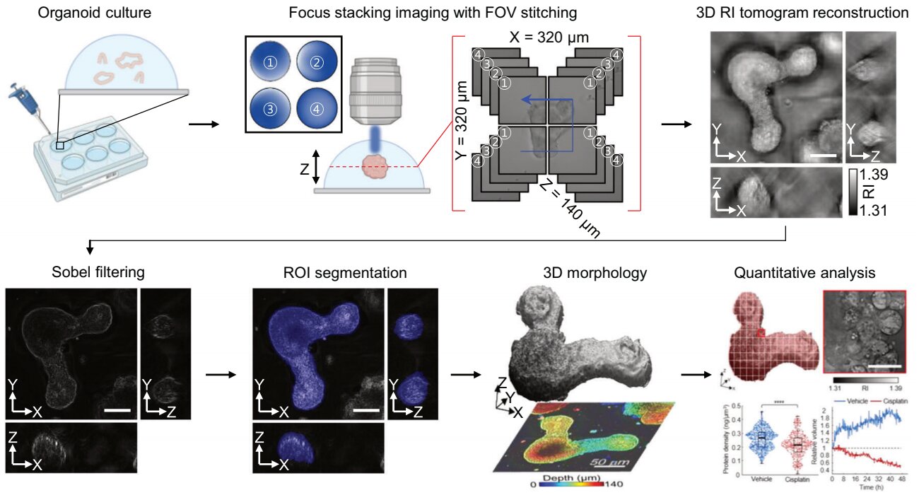
Overview of the low-consistency HT workflow. Using holotomography, 3D morphological restoration and quantitative analysis of organoids can be performed. In order to improve the limited field of view, which is a limitation of the microscope, our research team used a large area field of view combining algorithm and performed 3D restoration by acquiring multifocal holographic images for measurements. 3D. After that, the organoids were compartmentalized to divide the parts needed for analysis and quantitatively assessed the measurable protein concentration from the refractive index and survival rate of the organoids. Credit: Experimental and molecular medicine (2024). DOI: 10.1038/s12276-024-01312-0
Organoids, which are miniature 3D organs mimicking the structure and function of human organs, play a critical role in disease research and drug development. A Korean research team has overcome the limitations of existing imaging technologies and successfully observed living organoids in real time and at high resolution.
The research team of Professor YongKeun Park of the Department of Physics, together with the Genome Editing Research Center of the Institute of Basic Sciences and Tomocube Inc., developed imaging technology using holotomography to observe living small intestine organoids in real time. high resolution.
The research results were published in Experimental and molecular medicine on October 1, 2024, and the technology has been recognized for its applicability in various fields of life sciences.
Existing imaging techniques have struggled to observe living organoids in high resolution over long periods of time and have often required additional treatments like fluorescent staining.
The research team introduced holotomography technology to address these issues, which provides high-resolution images without fluorescent staining and allows long-term observation of dynamic changes in real time without damaging cells.
The team validated this technology using small intestinal organoids from experimental mice and were able to observe various cellular structures inside the organoids in detail. They also captured dynamic changes such as growth processes, cell division and cell death in real time using holotomography.
Analysis of organoid morphology in real time. Thanks to holotomography, it is possible to observe in real time the process of development of the lumen and villi of intestinal organoids, which was difficult to observe with a conventional microscope. Additionally, various information about intestinal organoids can be obtained by quantifying the size and protein quantity of intestinal organoids through image analysis. Credit: Experimental and molecular medicine (2024). DOI: 10.1038/s12276-024-01312-0
Additionally, the technology enabled the precise analysis of organoid responses to drug treatments, thereby verifying cell survival.
Researchers believe this breakthrough will open new horizons in organoid research, enabling greater use of organoids in drug development, personalized medicine and regenerative medicine.
Future research is expected to more accurately reproduce the in vivo environment of organoids, thereby significantly contributing to a more detailed understanding of various vital phenomena at the cellular level through more precise 3D imaging.
Dr. Mahn Jae Lee, a graduate of KAIST Graduate School of Medical Sciences and Engineering, currently at Chungnam National University Hospital and first author of the paper, commented: “This research represents a new technology of imaging that exceeds previous limitations and is expected to make a major contribution to disease modeling, personalized treatments and drug development research using organoids.
More information:
Mahn Jae Lee et al, Long-term three-dimensional high-resolution imaging of living unlabeled small intestinal organoids via low coherence holotomography, Experimental and molecular medicine (2024). DOI: 10.1038/s12276-024-01312-0
Provided by Korea Advanced Institute of Science and Technology (KAIST)
Quote: Holotomography allows real-time observation of organoids (October 15, 2024) retrieved October 15, 2024 from
This document is subject to copyright. Except for fair use for private study or research purposes, no part may be reproduced without written permission. The content is provided for informational purposes only.
