

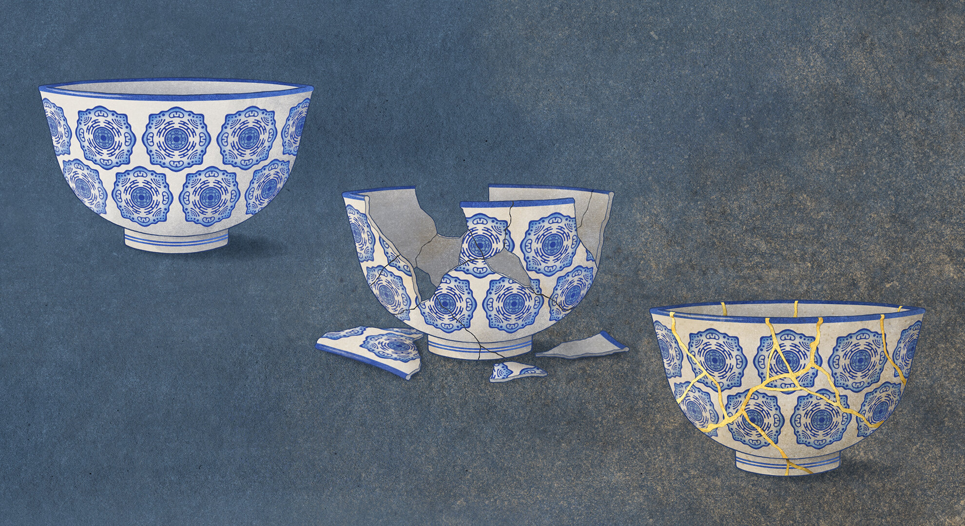
Kintsugi, the traditional Japanese art of repairing broken pottery by repairing the cracks with lacquer and gold. Kintsugi visibly integrates the history of an object into its new form. In this analogy, the damaged cell membrane is repaired. However, rather than restoring the cell to its original form, the new cellular nature is irreversibly changed and cells behave differently in our body. Credit: Amy Cao, Salk Institute
Our cells are surrounded by a fragile membrane only 5 nanometers thick, or 1/20 of a soap bubble. Cells are easily damaged by physiological activities, including muscle contraction and tissue damage. To cope with such damage, cells are equipped with mechanisms capable of repairing membrane damage to a certain extent.
Mechanical damage to the cell membrane was previously believed to trigger two simple cellular outcomes: healing or death. However, in a new study, researchers discovered a third outcome: cellular senescence. The work appears in Natural aging.
“When I started this project, my goal was simply to understand the repair mechanisms of the damaged cell membrane,” recalls Professor Keiko Kono, head of the Membrane Unit at the Institute of Science and Technology. Okinawa and lead author of the study, which included several members of the Okinawa Institute of Science and Technology. unit, including Kojiro Suda, Yohsuke Moriyama, Nurhanani Razali and colleagues. “Unexpectedly, we ended up discovering that damage to the cell membrane, in a sense, changes the fate of cells.”
The key to determining cell fate is the extent of damage and the subsequent influx of calcium ions. Damage to the thin cell membrane can be easily repaired, allowing cells to continue cell division without any problems. The highest level of cell membrane damage induces cell death. However, a medium level of cell membrane damage transforms cells into senescent cells several days later, even if membrane resealing appears successful.
Cancer cells divide unlimitedly. In contrast, normal non-cancerous cells have a limited capacity for cell division – about 50 times before division is irreversibly stopped and the cells enter a state called cellular senescence. Senescent cells are still metabolically active, but unlike young, healthy cells, they produce various secretory proteins that positively regulate immune responses in neighboring tissues and distant organs. This mechanism can induce both beneficial and harmful changes in our bodies, including accelerated wound healing, promotion of cancer, and aging.
Over the past decade, many studies have shown that senescent cells exist in the bodies of animals, including humans, and that removing senescent cells can rejuvenate the bodily functions of laboratory animals. However, the cause of cellular senescence in the human body remains a controversial topic.
“Gene expression profiling and bioinformatics suggest that cell membrane damage explains the origin of senescent cells in our body, especially those located near damaged tissues,” explains Professor Kono.
The best established inducer of cellular senescence is repeated cell division. Many other stresses also induce cellular senescence in the laboratory, such as DNA damage, oncogene activation, and epigenetic changes. The long-standing dogma in research has been that various stresses ultimately induce cellular senescence via activation of the DNA damage response.
However, the authors found that cell membrane damage induces cellular senescence via a different mechanism involving calcium ions and the tumor suppressor gene p53. These results could contribute to the development of a strategy to achieve healthy longevity in the future.
More information:
Damage to the plasma membrane limits the replicative lifespan of yeast and induces premature senescence of human fibroblasts, Natural aging (2024). DOI: 10.1038/s43587-024-00575-6
Provided by Okinawa Institute of Science and Technology
Quote: Damage to cell membranes causes cellular aging, according to a new study (February 22, 2024) retrieved February 22, 2024 from
This document is subject to copyright. Apart from fair use for private study or research purposes, no part may be reproduced without written permission. The content is provided for information only.
