

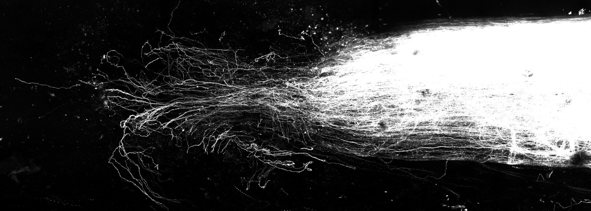
Imaging shows the regeneration of sensory axons four weeks after a spinal cord injury in an adult mouse injected with CEO-BB at the lesion site. Credit: Andrea Tedeschi / The Ohio State University
Capitalizing the flexibility of the tiny cells inside the smallest blood vessels in the body can be a powerful strategy for repairing spinal cord, suggests new research.
In mouse experiences, scientists introduced a specific type of recombinant protein on the site of a spinal cord lesion where these cells, called pericytes, had flooded the area of lesion. Once exposed to this protein, the results have shown that pericytes change their shape and inhibit the production of certain molecules while secreting others, creating “cell bridges” that support the regeneration of axons – long thin extensions of nerve cell body that transmit messages.
The researchers observed the regrowth of axons in injured mice which received a single injection of treatment of the growth factor protein, and the animals also found the movement in their posterior limbs. An experience involving human cells suggests that the results are not limited to mice.
“There is much more that can be learned and many things that can be widened, but the more we work there, the more we were really amazed by the power of this unique treatment and how effective it was,” said the author of the main study Andrea Tedeschi, associate professor of neuroscience at Ohio State University College of Medicine. “This observation goes beyond the lesions of the spinal cord – it has implications in brain lesions and cerebral vascular accidents, as well as neurodegenerative diseases.”
The work highlights the importance of restoring blood vessels to the recovery of neurological function after a spinal cord lesion, the researchers said.
“The lesions of the spinal cord are severe not only because they prevent the transmission of information on the injury site, but because the entire structure and function of the vascular system is also compromised,” said the first Wenjing Sun study, deputy professor of neuroscience at Ohio State. “Even if you are able to restore neural connectivity from one end to the other, the global effect will still not be maximized unless you take care of everything that collapses.”
The study is published in the journal Molecular therapy.
Previous research suggesting that pericytes interfere with the restoration of spinal cord lesions have led some scientists to recommend to eliminate them from the lesion site to facilitate repair. But cancer research has indicated that the properties of pericytes change when exposed to a protein called growth factor derived from BB plans (CEO -BB) – which is a way in which tumors generate their own blood supply. In cancer, the goal is to block PDGF-BB signaling.
Previous research on neuroscience has also indicated that pericytes are very “plastic”, which means that they are very sensitive to changes in microenvironnement, including the presence of CEO-BB. Tedeschi and his colleagues have seen a potential to exploit this cell protein relationship to stabilize the vascular system surrounding a spinal cord lesion. In the process, they found that the newly sprouted blood vessels had established a path to follow for the regenerated axons.
Starting with imaging studies, the team has shown that when a spinal cord is cut, pericytes migrate on the injury site over time but do not promote the growth of the functional blood vessels necessary to support the regeneration of axons.
In cell cultivation experiences, researchers have established a “carpet” of pericytes, added CEO, then placed a layer of adult mouse sensory neurons on top and evaluated the amount of axons increased in 24 hours. The treated axons have increased almost as much as healthy axons extend under normal conditions.
Pericytes, in blue and vascular, in pink, on the lesion site a month after a spinal cord injury in an adult mouse. Credit: Andrea Tedeschi / The Ohio State University
CEO alone did not produce this result. Instead, experiences have shown that pericytes combined with the growth factor rearranging fibronectin, a multifunctional adhesive glycoprotein which plays an essential role in the repair of tissues, the fixing of cells and motility. The cells themselves also change in shape, becoming more elongated.
“We know that these cells will infiltrate and deposit at the level of the epicenter of the lesion. These elongated fiber structures that they become are much more permissive to promote the axons to regenerate from one end to the other and bypass the injury,” said Tedeschi.
“To extend the clinical relevance of our results, we have cultivated mouse neurons above human pericytes which were exposed to the CEO-BB, and which was sufficient to trigger an effect promoting growth, suggesting that this could really be a generalized phenomenon which is not limited to mouse.”
Turning to experiences in animals with spinal cord lesions, the researchers waited seven days after the injury – the equivalent of about nine months in a human adult – injecting before a single dose of PDGF -BB on the injury site. The tissue analysis four weeks after the injury showed that the injection of CEO-BB produced regenerative growth of the robust axons compared to the Axone response in wounded control mice.
“When we examined the formation of these pericyte structures that crossed the injury site, we saw the treatment favored the growth of these bridges. And most, if not all of these regenerating axons, were able to escape the injury site by driving these cell bridges which were trained in response to the PDGF-BB administration,” said Sun.
The electrophysiological and movement evaluations of injured animals treated with PDGF-BB have detected sensory activity beyond the lesion site and have shown that mice have regained better control of their posterior members compared to witness mice. The animals were also less sensitive to an unpainted stimulus, suggesting that they had not felt the neuropathic pain which is often triggered by a spinal cord lesion.
Analysis of the presence of inflammatory proteins during the repair process suggested that the administration of PDGF-BB not only promotes the regeneration of axons, but also reduces inflammation. The sequencing of RNA has shown that the lesion of the spinal cord has led to a decrease in the expression of genes by pericytes, but that the cells have retained their central properties and have not turned into a different cell type – for example, a type of cell that could end up being destructive for the injury environment.
“There has been a decrease in certain markers of conventional pericytes, but a gain of an additional function linked to the attempt to reconstruct cell bridges and functional vessels,” said Sun. “According to the overall signature of the genes of our data, they are always classified as pericyte.”
Since Tedeschi, Sun and his colleagues have previously shown in mice that gabapentin promotes the regeneration of neural circuits after a spinal cord lesion, it is possible to consider a multi-plaster approach to therapy, said Sun.
“We could combine the two-modulating the intrinsic properties of adult neurons with a drug and what we do here, modulating the non-neuronal environment to produce cellular interactions that provide a more permissive substrate so that the neuron is developing,” she said.
More work is planned to determine the precise moment for the administration of CEO -BB – with the presumption that the pericytes take a while to migrate to the injury – as well as the ideal concentration of treatment and a potential delivery system on time.
More information:
Wenjing Sun et al, in vivo programming for adult pericytes helping the regeneration of axons by providing cell bridges for the repair of SCI, Molecular therapy (2025). DOI: 10.1016 / J.YMTHE.2025.04.020
Supplied by the Ohio State University
Quote: Build “cell bridges” for repairing the spinal cord after an injury (2025, April 21) recovered on April 21, 2025
This document is subject to copyright. In addition to any fair program for private or research purposes, no part can be reproduced without written authorization. The content is provided only for information purposes.