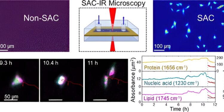
Credit: Analytical chemistry (2024). DOI: 10.1021/acs.analchem.4c02108
To accelerate biotechnological innovations, such as the development of life-saving drug therapies, scientists are working to develop faster, more quantitative, and more widely available ways to observe biomolecules in living cells.
Researchers at the National Institute of Standards and Technology (NIST) have developed a new method that uses infrared (IR) light to capture clear images of biomolecules inside cells, something that was not previously possible because of the tendency of the water in cells to absorb infrared radiation. Their findings were published in Analytical chemistry.
This new method eliminates the obscuring effects of water in infrared measurements and allows researchers to determine the quantities of key biomolecules in cells, such as proteins that direct cellular function. The ability to measure changes in living cells could accelerate advances in biomanufacturing, cell therapy development, drug development and much more.
Infrared radiation is light that is just beyond what is visible to the human eye. Although we cannot see infrared light, we can sense it as heat. In infrared microscopy, a material of interest absorbs radiation from a range of wavelengths in the infrared spectrum.
Scientists measure and analyze the infrared absorption spectrum of a sample, producing a set of “fingerprints” that can identify molecules and other chemical structures. However, water, the most abundant molecule inside and outside cells, strongly absorbs infrared and masks the infrared absorption of other biomolecules in the cell.
One way to understand this optical masking effect is to compare it to a plane flying overhead, next to the sun. With the naked eye, it is difficult to see the plane because of the sun, but if you use a special sun-blocking filter, you can easily see the plane in the sky.
“In the spectrum, water absorbs infrared very strongly, and we want to see the absorption spectrum of proteins through the thick background of water, so we designed the optical system to unveil the contribution of water and reveal the signals from proteins,” said NIST chemist Young Jong Lee.
Lee developed a patented technique that uses an optical element to compensate for the absorption of water by infrared. Called solvent absorption compensation (SAC), this technique was used with a hand-built IR laser microscope to image the cells that support the formation of connective tissue, called fibroblast cells.
During a 12-hour observation period, the researchers were able to identify groups of biomolecules (proteins, lipids and nucleic acids) during different stages of the cell cycle, such as cell division. Although this time may seem long, the method is ultimately faster than current alternatives, which require beam time in a large synchrotron facility.
This new method, called SAC-IR, is label-free, meaning it does not require any dyes or fluorescent labels, which can damage cells and also produce less consistent results from lab to lab.
The SAC-IR method allowed NIST researchers to measure the absolute mass of proteins in a cell, in addition to nucleic acids, lipids and carbohydrates. This technique could help lay the foundation for standardizing methods for measuring biomolecules in cells, which could prove useful in biology, medicine and biotechnology.
“In cancer cell therapy, for example, when cells from a patient’s immune system are modified to better recognize and kill cancer cells before being reintroduced into the patient’s body, there are questions about whether these cells are safe and effective. Our method can be useful by providing additional information about biomolecular changes in cells to assess their health,” Lee said.
Other potential applications include using cells for drug screening, either for the discovery of new drugs or to understand the safety and efficacy of a drug candidate. For example, this method could help assess the efficacy of new drugs by measuring the absolute concentrations of various biomolecules in large numbers of individual cells, or to analyze how different cell types respond to drugs.
The researchers hope to further develop this technique so that they can more accurately measure other key biomolecules, such as DNA and RNA. The technique could also help provide detailed answers to fundamental questions in cell biology, such as how biomolecule signatures correspond to cell viability—whether a cell is alive, dying, or dead.
“Some cells are stored frozen for months or years and then thawed for later use. We don’t yet fully understand the best way to thaw cells while maintaining maximum viability. With our new measurement capabilities, we may be able to develop better processes for freezing and thawing cells by looking at their infrared spectra,” Lee said.
More information:
Yow-Ren Chang et al., Benchtop IR imaging of live cells: monitoring the total mass of biomolecules in individual cells, Analytical chemistry (2024). DOI: 10.1021/acs.analchem.4c02108
Provided by the National Institute of Standards and Technology
This article is republished with kind permission from NIST. Read the original article here.
Quote:Biomolecules inside living cells can now be observed in infrared light using a new method (2024, September 9) retrieved September 9, 2024 from
This document is subject to copyright. Apart from any fair dealing for the purpose of private study or research, no part may be reproduced without written permission. The content is provided for informational purposes only.


