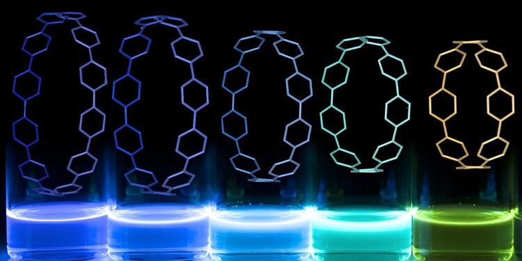
The ring-shaped “nanohoops” emit different colors of light depending on their structure. Credit: University of Oregon
In a process as simple as mixing eggs and flour into pancakes, researchers at the University of Oregon mixed ring-shaped fluorescent molecules in a new 3D printing process. The result: complex light structures that support the development of new types of biomedical implants.
This advancement solves a long-standing design challenge by making structures easier to track and monitor over time inside the body, allowing researchers to easily distinguish what is part of an implant and what is constitutes cells or tissues.
The discovery is the result of a collaboration between Paul Dalton’s on-campus engineering lab Phil and Penny Knight to Accelerate Scientific Impact and Ramesh Jasti’s chemistry lab in the OU College of Arts and Sciences . The researchers describe their findings in a paper published this summer in the journal Little.
“I think it was one of those weird moments where we said, ‘Let’s try,’ and it worked almost immediately,” Dalton said.
But behind this simple origin story lie years of specialized research and expertise in two very different fields before they finally came together.
Dalton’s lab specializes in new and complex forms of 3D printing. His team’s flagship development is a technique called fusion electrowriting, which allows relatively large objects to be 3D printed with very fine resolution. Using this technique, the team printed mesh scaffolds that could be used for different types of biomedical implants.
Fusion electrowriting is a new 3D printing technique developed by Dalton. Credit: University of Oregon
Such implants could be used for applications as diverse as new wound healing technologies, artificial blood vessels or structures helping to regenerate nerves. In a recent project, the lab collaborated with cosmetics company L’Oréal, using the scaffolding to create realistic, multi-layered artificial skin.
Jasti’s lab, meanwhile, is known for its work on nanohoops, ring-shaped carbon-based molecules that have a variety of interesting properties and are tunable based on the precise size and structure of the hoops in ring shape. The nanohoops fluoresce intensely when exposed to ultraviolet light, emitting different colors depending on their size and structure.
The two labs might have stayed in their own lanes without a casual conversation when Dalton was a new professor at OU, eager to make connections and meet other faculty members. He and Jasti floated the idea of incorporating the nanohoops into the 3D scaffolds Dalton was already working on. This would make the structures glow, a useful feature that would make it easier to track their fate in the body and distinguish structures from their surroundings.
“We thought it probably wouldn’t work,” Jasti said. But it did, quite quickly.
People had tried to shine the scaffolding in the past, without much success, Dalton said. Most fluorescent molecules break down under the long exposure to heat required for his 3D printing technique. The nanohoops from the Jasti laboratory are much more stable at high temperatures.
The nano hoops glow under UV light. Credit: University of Oregon
Although both groups may make their craft seem easy, “making nanohoops is really hard, and fusion electrowriting is really hard to do, so the fact that we were able to merge these two really complex fields and different into something really simple is amazing,” said Harrison Reid, a graduate student in Jasti’s lab.
According to the researchers, a small amount of fluorescent nanohoops mixed into the mixture of 3D printing materials produces long-lasting glowing structures. Because fluorescence is activated by UV light, the scaffolds still appear clear under normal conditions.
Although the initial concept worked very quickly, it took several years of additional testing to fully evaluate the material and gauge its potential, said Patrick Hall, a graduate student in Dalton’s lab.
For example, Hall and Dalton performed a battery of tests to confirm that adding nanohoops did not affect the strength or stability of the 3D printed material. They also confirmed that the addition of fluorescent molecules did not make the resulting material toxic to cells, which is important for biomedical applications and is a key baseline that must be met before it can get any closer to application. human.
The team envisions a range of possible applications for the luminous materials they created. Dalton is particularly interested in the biomedical potential, but a customizable material that glows under UV light could also be used in security applications, Jasti said.
A close-up of a scaffold made of nanohoops, glowing blue under UV light. Credit: University of Oregon
They have filed a patent application for this advancement and hope to eventually commercialize it. And both Jasti and Dalton are grateful for the chance that brought them together.
“We get interesting new directions by bringing together people who don’t typically discuss their science,” Dalton said.
More information:
Patrick C. Hall et al, (n) Cycloparaphenylenes as compatible fluorophores for fusion electrowriting, Little (2024). DOI: 10.1002/smll.202400882
Journal information:
Little
Provided by the University of Oregon
Quote: Bioengineers and chemists design 3D printed fluorescent structures with potential medical applications (September 27, 2024) retrieved September 27, 2024 from
This document is subject to copyright. Except for fair use for private study or research purposes, no part may be reproduced without written permission. The content is provided for informational purposes only.


