

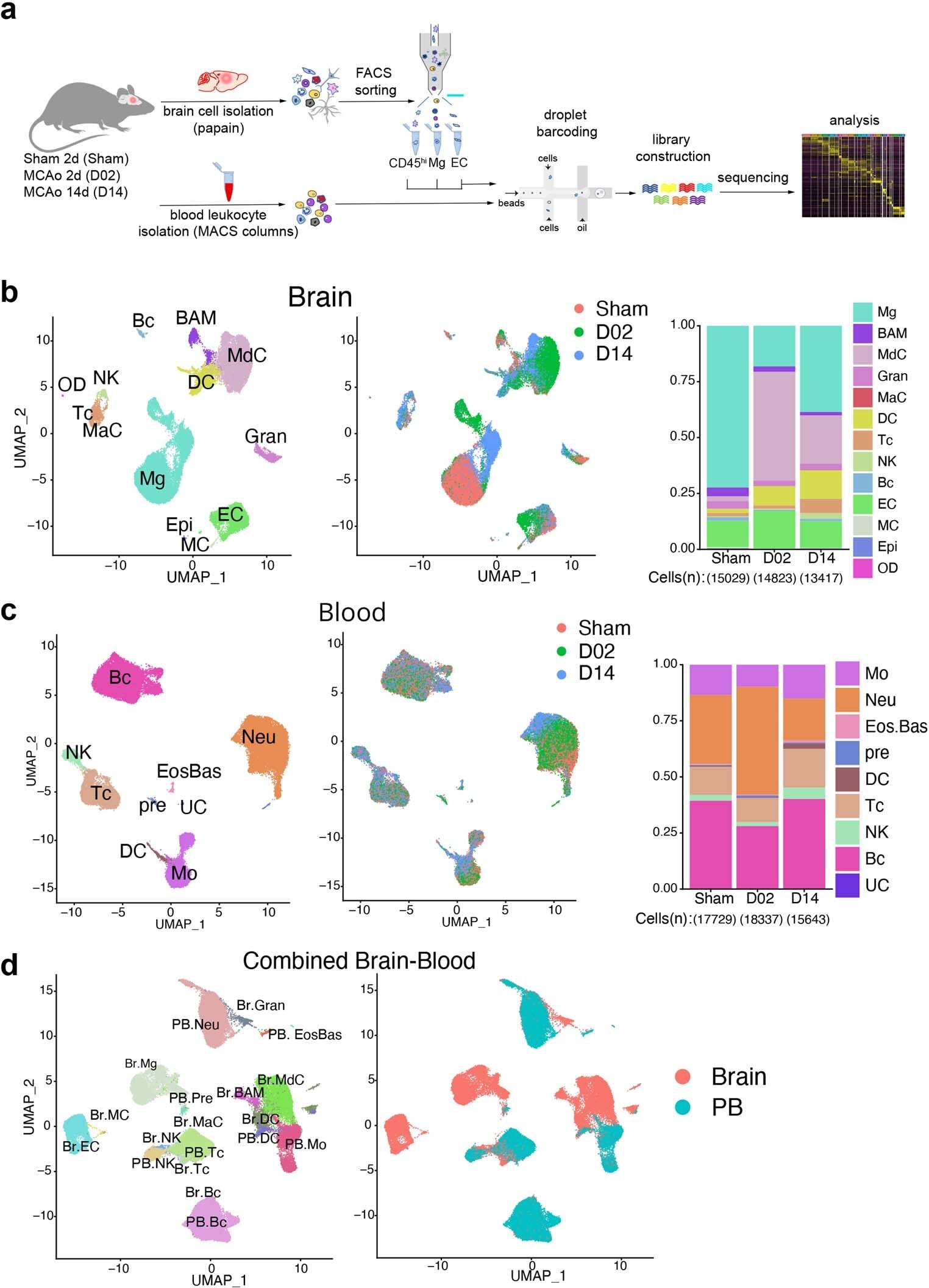
Single-cell transcriptomic profiling of mouse brain and blood cells after transient focal cerebral ischemia. Credit: Natural immunology (2024). DOI: 10.1038/s41590-023-01711-x
Weill Cornell Medicine researchers have cataloged the cellular response to stroke in a preclinical model, identifying the immune cells involved and the roles they may play in the days and weeks following a stroke.
During a stroke, loss of oxygen leads to brain damage and cell death. It also triggers a potent inflammatory response in which resident immune cells in the brain, as well as cells recruited from the blood, infiltrate injured tissue.
The results, published on January 4 in Natural immunologycould point to new approaches to promote recovery from stroke and provide insight into why therapies aimed at controlling inflammation after stroke have not been successful.
“Almost every one of us knows someone who has had a stroke. It’s a huge problem,” said lead author Dr. Josef Anrather, professor of neuroscience and vice president for research at the Feil Family Brain and Mind Research Institute at Weill Cornell Medicine. “But in terms of treatment, there is only so much a doctor can do.”
Interventions that restore blood flow to the affected brain region must be administered within a few hours to be effective. “So most people, over 80 percent, don’t get any therapy,” he said.
Understanding how immune cells help repair and remodel the brain during the later chronic phase after a stroke could help doctors minimize long-term neurological consequences, including dementia and even seizures.
In 2016, Anrather and colleagues observed that immune cells called monocytes, made in the bone marrow, accumulated in the brain following a stroke. Once there, they appeared to undergo a physical transformation: some grew spindly arms, adopting the appearance of the brain’s resident immune cells, microglia; others became more amorphous and amoeba-like.
But what, if anything, did this shapeshift have to do with their behavior?
“We were interested in knowing the function of these different structural features,” said the study’s lead author, Lidia Garcia-Bonilla, a Finbar and Marianne Kenny Scholar in Neurology and research assistant professor of neuroscience at Brain and Mind. Weill Research Institute. Cornell Medicine.
They also wondered whether these cells contributed to recovery or worsened the damage.
“There are always two sides to the coin,” Anrather said. The same type of cell can be harmful in some circumstances but useful in others. “This may be why clinical trials of drugs that reduce brain immune cell infiltration and inflammation have shown no benefit in stroke.”
The most direct way to assess what a particular cell is doing is to determine which of its many genes are turned on. Working with a preclinical model, they collected immune cells two days and 14 days after an induced stroke – the blockage of an artery in the brain. They then sequenced the RNA molecules, which code for proteins, produced by each cell. Using this approach, the researchers identified exactly each cell type they had isolated. It also showed which genes each cell had activated, an indication of their role after the stroke.
The researchers first noticed that a population of microglia was rapidly proliferating. This made sense, Anrather said, “because microglia cover the territory of the brain.” When their numbers decrease due to an injury, such as a stroke, the cells multiply to cover damaged tissue.
Then they “take out the trash,” Anrather said.
“For the brain to rebuild itself, you have to clean, eliminate dead cells,” he explained. Indeed, two days after the experimental stroke, the researchers detected a cadre of microglia that activate genes involved in the removal of cellular debris.
Monocytes, white blood cells that responded to injury, joined microglia in this effort. “These cells are continually circulating and don’t really have a job until there’s a problem, like infection, trauma or any sort of tissue death,” Anrather said. “Then they’re called in to help clean up.”
Once there, the researchers found, these monocytes transformed into the type of cell needed to get the job done. “They are like little children who are educated in the fabrics,” Anrather said.
After the acute cleansing phase, the immune response was restructured towards tissue remodeling. Some cellular recruits produced repair-triggering growth factors while immunological “professionals” such as T cells were called upon to play a neuroprotective role.
By identifying which immune cells will respond to the stroke-induced distress call, researchers provide a new vehicle for intervention. “Because these cells know how to reach the brain,” Anrather said, “you could use them as a shuttle and engineer them to deliver treatment.”
Additionally, understanding precisely what these cells do when they reach the brain could be key to developing treatments that can be administered weeks or months after a stroke. “Finding a way to activate the brain’s natural repair mechanism could improve outcomes for stroke patients,” Garcia-Bonilla said.
More information:
Lidia Garcia-Bonilla et al, Analysis of single-cell transcriptomics of brain and blood in the acute and subacute phases after experimental stroke, Natural immunology (2024). DOI: 10.1038/s41590-023-01711-x
Provided by Cornell University
Quote: Mapping brain repair and remodeling after stroke (January 10, 2024) retrieved January 10, 2024 from
This document is subject to copyright. Apart from fair use for private study or research purposes, no part may be reproduced without written permission. The content is provided for information only.
