

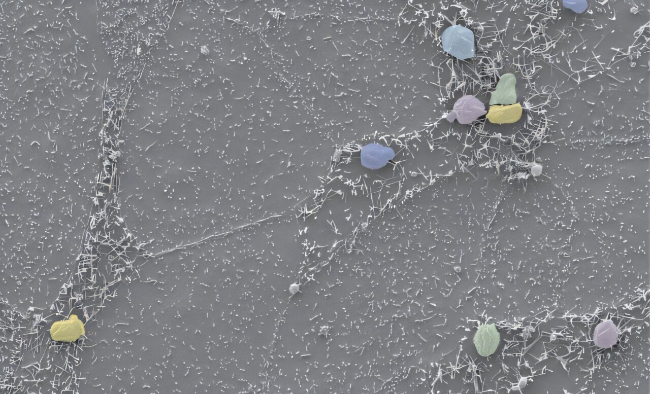
Small spherical vacuoles (colored spheres) which shelter the cryptosporidium parasite can be easily detected on the cell surface. Here, the parasite exports proteins to the host cell that handle function and appearance, including the lengthening of microvillis which line the intestinal surface (longer gray strands). Credit: Elena Rodrigues
Researchers from the Francis Crick Institute have shown that the Cryptosporidium parasite exports a protein to infected intestinal cells, modifying the intestinal environment and allowing the parasite to survive and reply.
Cryptosporidium is an intestinal parasite and one of the main causes of severe diarrhea in children. It invades and replies inside the epithelial cells which line the intestine from the host and is known to cause projections absorbing nutrients called microvillis.
In the research published in Cell host and microbeThe Crick laboratory identified a family of protein that Cryptosporidium injects in intestinal epithelial cells during an infection, which accumulates in the host microvilloscopic.
The team studied a major protein in this family, called microvilli protein 1 (MVP1), noting that, in the epithelial cell, it interacts with human proteins which are responsible for maintaining the structure and regulation of cellular projections such as microvilis.
When MVP1 was removed from a strain of cryptosporidium, using genetic edition based on CRISPR, lengthening the microville in infected cells was completely blocked. This strain of deficient parasites in MVP1 has not caused normal infection in mice either.
One of the proteins with which MVP1 interacts, called EBP50, is crucial to stabilize the pumps on the surface of cells which bring salts. The disturbance of these pumps leads to diarrhea, so that the team thinks that MVP1 could be one of the key factors that lead the symptoms caused by cryptosporidium.
Adam Steriale, group leader of the cryptosporidiosis laboratory of Crick, said: “The disease caused by Cryptosporidium is particularly dangerous for children and it can cause hard malnutrition to prevent lasting diseases and the resulting malnutrition.”
The team compared the exported proteins of cryptosporidium and E. coli bacteria. Credit: Niaid
Convergent evolution: similar mechanisms in bacteria and parasites
Cryptosporidium is not the only pathogen known to cause an extension of microvilli in the intestine. A type of e. Coli also infects the intestine and causes diarrhea. This type of e. Coli is also known to stimulate the extension of microvillis, by the action of a protein called map which manipulates structural proteins in intestinal epithelial cells during an infection.
Surprisingly, the team noted that MVP1 of Cryptosporidium interacts with the same structural proteins as the card of E. Coli, EBP50 and CDC42. The team believes that this is a unique occurrence of a convergent evolution, where proteins causing a disease in bacteria and infectious parasites have evolved independently to have the same function.
Elena Rodrigues, former doctorate. The student in the cryptosporidiosis laboratory of Crick, said: “The similarities of the effective proteins between a type of E. coli and cryptosporidium show a fascinating example of convergent evolution: how the pathogens of two different kingdoms have all the needs of the advanced protein separate foundation.”
More information:
Elena Rodrigues et al, cryptosporidium modifies microvillos Cell host and microbe (2025). DOI: 10.1016 / J.CHOM.2025.04.001
Provided by the Francis Crick Institute
Quote: Researchers discover how the Cryptosporidium Intestinal Parasite modifies host cells (2025, April 28) recovered on April 28, 2025 from
This document is subject to copyright. In addition to any fair program for private or research purposes, no part can be reproduced without written authorization. The content is provided only for information purposes.
