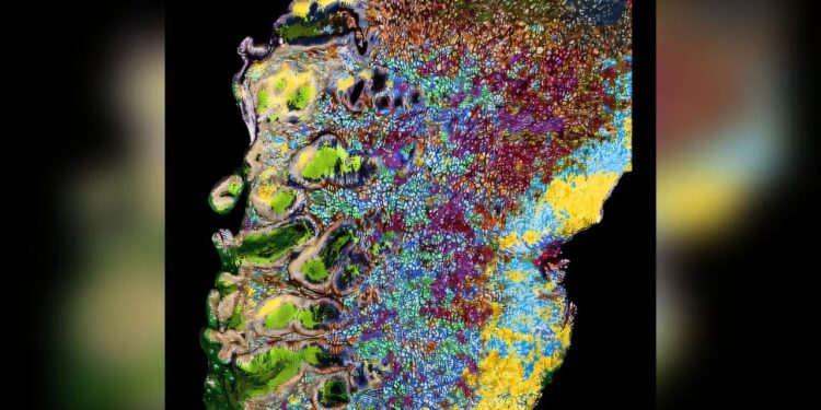
Patho-DBiT reveals tissue architecture at the cellular level of an aggressive gastric lymphoma sample stored for three years. Credit: Yale University
A new pathology tool created at Yale leverages barcode technology and shows its potential for use in cancer diagnostics. The technology, Patho-DBiT (deterministic barcode compatible with pathology in tissues), was discussed in a new study published in the journal Cell.
Co-corresponding author Mina Xu, MD, professor of pathology and laboratory medicine, member of the Yale Cancer Center (YCC), and director of hematopathology at YSM, shared her enthusiasm for the new tool.
“As a doctor who diagnosed cancer, I was surprised how much more I can see using this pathology tool,” Xu said. “I believe this molecular deep dive will advance our understanding of tumor biology exponentially. I really look forward to providing more precise and actionable diagnostics.”
Patho-DBiT uses DNA barcoding to map the spatial relationships between RNA and proteins, allowing comprehensive examination of RNA (some types play regulatory roles in cancer). The technology is unique in that it features microfluidic devices that deliver barcodes into tissue from two directions, creating a unique 2D “mosaic” of pixels, providing spatial information that could be used to inform the creation of patient-specific targeted therapies. The technology, created in the lab of Yale Ph.D. Rong Fan, is now licensed to AtlasXomics, a Yale spinoff company.
“This is the first time we can directly ‘see’ all kinds of RNA species, where they are and what they do, in clinical tissue samples,” said Fan, Harold Hodgkinson Professor of biomedical engineering and pathology at YSM, lead author of the study. study and member of the YCC. “With this tool, we are able to better understand the fascinating biology of each RNA molecule that has a very rich life cycle, beyond just knowing whether each gene is expressed or not. I think that “This will completely transform the way we study human biology in the future.”
In the study manuscript, the researchers explain why their tool, Patho-DBiT, could be the key to unlocking a “wealth of information” preserved in laboratory tissue biopsy samples.
“Millions of these tissues have been archived for so many years, but until now we did not have effective tools to study them at the spatial level,” said the study’s first author, Zhiliang Bai, Ph.D., researcher. postdoctoral associate in Fan’s lab. “The RNA molecules in the tissues we study are highly fragmented and traditional methods cannot capture all the important information about them. This is why we are very excited about Patho-DBiT.”
Potential future uses of Patho-DBiT include creating targeted therapies and helping to understand the mechanism of how low-grade tumors transform into more aggressive tumors in order to find ways to prevent it. For Patho-DBiT to be included in pathological diagnostics, researchers say more studies are needed to test and validate patient samples.
This multidisciplinary research included faculty members from several departments at Yale, including biomedical engineering, pathology, and genetics. Jun Lu, Ph.D., associate professor of genetics, joined Fan, Bai and Xu as a co-author from Yale.
“It is very interesting that Patho-DBiT-seq is also able to generate spatial maps of the expression of non-coding RNAs,” said Lu. “Non-coding RNAs are often found in regions of our genome that were previously previously considered junk DNA, but are now recognized as valuable players in biology and in diseases such as cancer.”
More information:
Zhiliang Bai et al, Spatial exploration of RNA biology in formalin-fixed and paraffin-embedded archival tissues, Cell (2024). DOI: 10.1016/j.cell.2024.09.001
Cell
Provided by Yale University
Quote: New barcode technology could help diagnose cancer more accurately (November 20, 2024) retrieved November 20, 2024 from
This document is subject to copyright. Except for fair use for private study or research purposes, no part may be reproduced without written permission. The content is provided for informational purposes only.


