

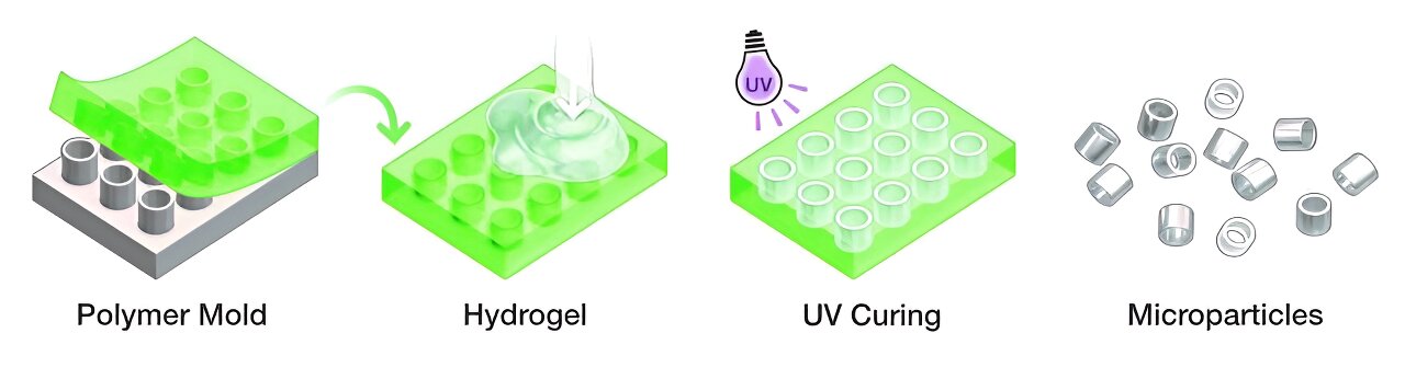
A hard silicone master mold is used to create a flexible polymer mold, which is inverted and filled with hydrogel. The hydrogel is then cured under UV light, producing the cylindrical microparticles. Credit: S. Kelley/NIST
Microscopic magnetic probes that change shape in response to their environment can greatly improve magnetic resonance imaging (MRI). However, the production of these probes, still experimental and not yet used in humans, required access to a clean room and expertise in nanofabrication, limiting their widespread use.
Now, researchers at the National Institute of Standards and Technology (NIST) have taken these shape-changing probes, known as geometrically encoded magnetic sensors, or GEMS, a step further by unveiling a new method manufacturing which is not only faster and cheaper, but eliminates the need for specialized instrumentation.
The scientists reported their work online on December 19 in ACS sensors.
Instead of building the tiny probes layer by layer in a nanofabrication facility, the team built them using a precision master mold. This technique allows researchers to make GEMS in their own laboratories using inexpensive materials and readily available equipment.
NIST scientists Gary Zabow and Samuel Oberdick and their colleagues focused their efforts on building GEMS in the shape of tiny hollow cylinders, because this shape can be easily made with a mold. For their main mold, the scientists constructed a set of hollow cylinders made of hard silicon, each measuring only about 100 micrometers in diameter, or about ten times larger than a red blood cell.
The microparticles are soaked in a bath of iron salts, impregnating the hydrogel with iron salts. They are then transferred to a high pH solution, which transforms the iron salts contained in the hydrogel into magnetic iron oxide particles. Credit: S. Kelley/NIST
The team then demonstrated how researchers with such a master mold could carry out the multi-step manufacturing process. First, the scientists made a “negative” of the soft mold by pouring a liquid polymer onto the hard silicone mold, allowing it to solidify, and then peeling it off. This created a collapsible mold with a set of cylindrical hollow cavities.
In the next step, the scientists filled each cavity with a liquid precursor to a hydrogel, a network of cross-linked polymers capable of absorbing large amounts of water. A key component of GEMS is the hydrogel, which has been designed to shrink or swell in response to changes in acidity or other properties of its microenvironment. Engineered hydrogels are inexpensive and easy to manufacture.
After hardening the hydrogels by exposing them to ultraviolet light, the NIST team took them out of their soft mold, much like taking ice cubes out of a silicone tray. The cylindrical hydrogels were then immersed in a bath of iron salts and transferred to a basic solution, which converted the iron salts absorbed by the hydrogels into magnetic oxide particles.
The strength of each hydrogel’s magnetic field directly impacts the MRI, which manipulates the tiny magnetic fields of protons to image the human body’s internal structures. Protons behave like rotating magnetized tops, each initially pointing in a random direction.
When placed in a strong external magnetic field (labeled M), iron oxide particles become magnetized, causing the microparticles to develop their own local magnetic field. The microparticles shrink and swell with changes in acidity, which strengthens or weakens this local field and therefore how much this field influences the proton resonance frequency in an MRI scan. Credit: S. Kelley/NIST
An MRI machine aligns the protons’ magnetic field with its own strong magnetic field, then disrupts that alignment by tickling the protons with a pulse of radio waves at a resonant frequency that causes the protons to alternately “relax” into their original state. origin, then become aligned again. As the protons shuttle between the two states, they emit radio waves, which are translated into MRI images.
During this time, the hydrogels change shape in response to changes in local conditions, leading to a strengthening or weakening of their magnetic field.
The changing magnetic field of GEMS shifts the resonance frequency of protons that are in or near the probes. By measuring this change, MRI can detect how GEMS have changed their shape in response to a specific property of their local environment.
GEMS built with the soft casting process can be tailored to change their shape based on a multitude of environmental properties, allowing researchers to use the probes to explore a range of biomedical conditions, Oberdick said.
More information:
Samuel D. Oberdick et al, Shaped magnetogel microparticles for multispectral magnetic resonance contrast and detection, ACS sensors (2023). DOI: 10.1021/acsensors.3c01373
Provided by National Institute of Standards and Technology
This story is republished courtesy of NIST. Read the original story here.
Quote: Ultra-small, shape-shifting GEMS offer a simpler, cheaper way to improve MRI imaging (December 20, 2023) retrieved December 21, 2023 from
This document is subject to copyright. Apart from fair use for private study or research purposes, no part may be reproduced without written permission. The content is provided for information only.
