

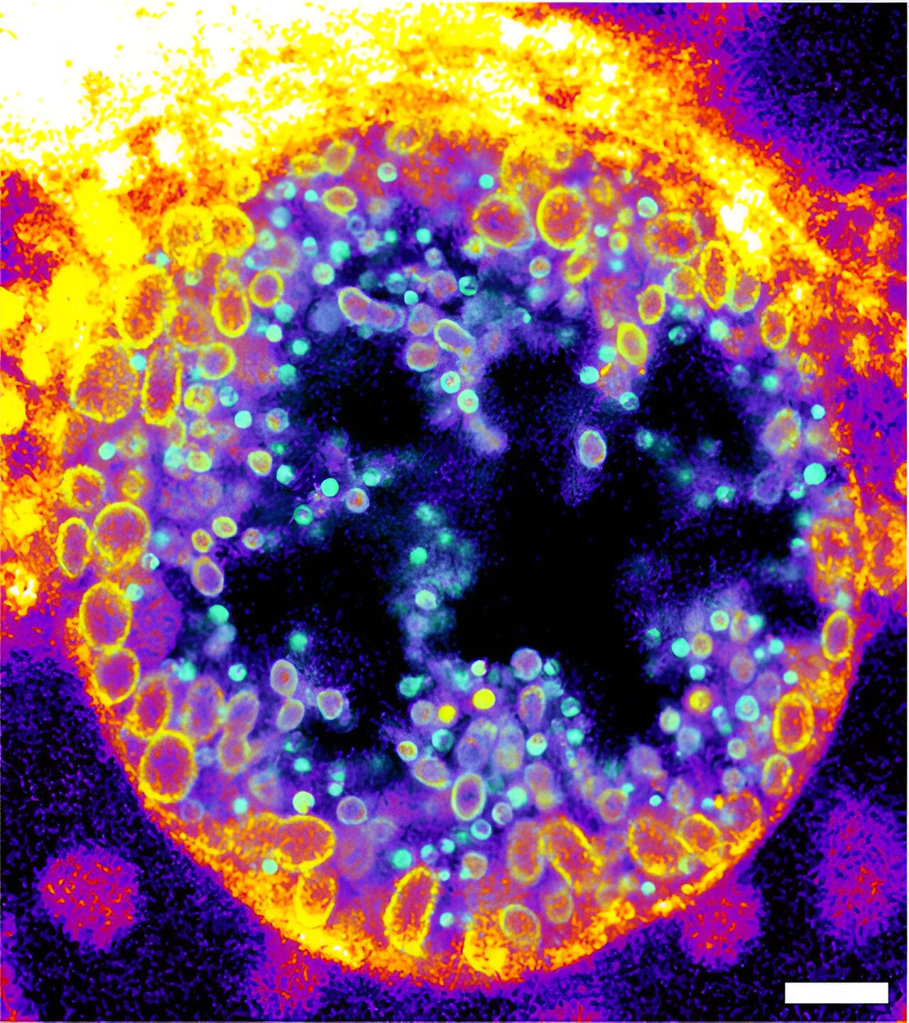
Novel trifunctional sphingomyelins (TFSMs) can be used to visualize chlamydial inclusions in infected human cells and allow the detection of native sphingomyelin derivatives in noninfectious reticulate bodies (yellow circles) and metabolized sphingomyelin derivatives in infectious elementary bodies (green dots). Credit: Jürgen Seibel / University of Würzburg
Researchers from Würzburg and Berlin present a new molecule that can visualize the metabolism of sphingomyelin. This opens up perspectives for innovative therapeutic approaches in infection research. The work is published in the journal Nature Communications.
In the late 19th century, German pathologist Ludwig Thudichum isolated previously unknown fatty substances (lipids) from the brain. He named this new class of molecules sphingolipids, after the Greek mythical creature the Sphinx, out of respect for “the many puzzles it posed to researchers.”
Since then, many diseases caused by defective sphingolipid metabolism in the brain have been discovered, including Fabry disease and Gaucher disease. Sphingolipids have also been linked to infectious diseases, for example viral infections such as Ebola, measles or COVID-19, as well as bacterial infections such as Pseudomonas aeruginosa or Staphylococcus aureus, which can cause infections of the middle ear, skin and lungs, as well as many other diseases.
In these infections, the degradation of the sphingomyelin molecule by the enzyme sphingomyelinase is often a crucial step. However, it was until now impossible to visualize the activity of the enzyme.
New chemical probe to fill the gap
Researchers from Würzburg and Berlin have now succeeded in developing a sphingomyelin derivative that allows the distribution of sphingomyelin and the activity of sphingomyelinase to be visualized in infection processes.
The scientists are part of Research Training Group 2581 “Metabolism, Topology and Compartmentalization of Membrane-Proximal Lipid and Signaling Components in Infection.” Chemists, physicists and biologists collaborated to synthesize new chemical compounds and test their applicability in infection research.
“The new molecules are trifunctional sphingomyelins based on the natural product sphingomyelin and with three additional functions. It was difficult to design molecules that are accepted by the metabolism as their natural origin,” explains Professor Jürgen Seibel from the Institute of Organic Chemistry at the Julius-Maximilians-University (JMU) Würzburg in Bavaria, Germany.
Imaging sphingomyelin degradation during chlamydia bacterial development
The scientists demonstrated the function of their newly developed molecules not only by determining the activity of a bacterial sphingomyelinase on the surface of human cells, but they also visualized the degradation of sphingomyelin in human cells during infection with intracellular Chlamydia bacteria, which are known to infect the human genital tract and are suspected of contributing to the development of cancer in infected tissues.
Within their host cells, chlamydia form a replicative organelle called an inclusion. Researchers have shown that chlamydial inclusions contain mainly the cleaved forms of trifunctional sphingomyelins.
Using expansion microscopy and click-chemistry, they observed that the proportion of metabolized sphingomyelin molecules increased during the maturation of non-infectious to infectious Chlamydia particles. Visualizing this infection process now allows the development of new targeted strategies against these infections.
“This new chemical tool will certainly be useful to us and can be used in many laboratories,” says Professor Seibel. “Our goal is to use it to identify new anti-infective or immunotherapeutic strategies for the development of drugs that can be used to combat infectious diseases by modulating sphingolipid metabolism.”
More information:
Marcel Rühling et al, Trifunctional sphingomyelin derivatives enable nanoscale resolution of sphingomyelin turnover in physiological and infectious processes via expansion microscopy, Nature Communications (2024). DOI: 10.1038/s41467-024-51874-w
Provided by Julius-Maximilians-Universität Würzburg
Quote:Scientists develop new chemical tool for infection research (2024, August 29) retrieved August 29, 2024 from
This document is subject to copyright. Apart from any fair dealing for the purpose of private study or research, no part may be reproduced without written permission. The content is provided for informational purposes only.
