

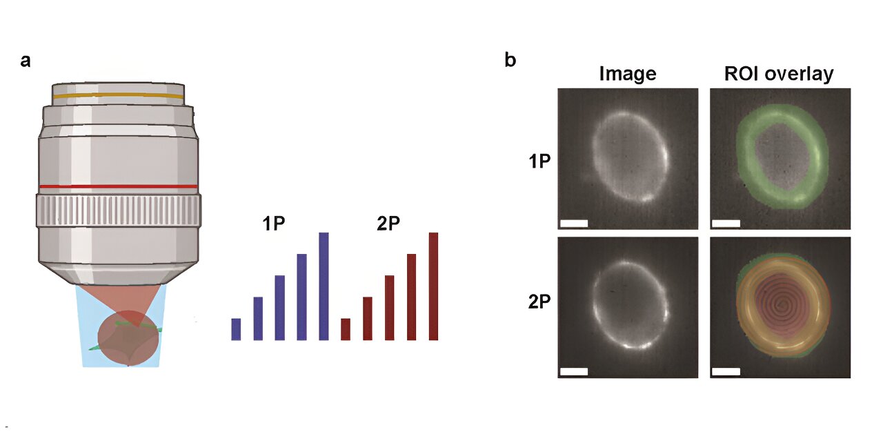
Comparison of the brightness and sensitivity of one-photon (1P) and two-photon (2P) fluorescent voltage indicators. (a) Embryonic kidney cells were sequentially illuminated with wide-field 1P light in increasing intensity steps, followed by spiral scanning in increasing intensity steps at 2P. (b) Example of a cell expressing a genetically encoded voltage indicator. The 2P spiral scanning pattern is shown in red and the region of interest scan is shown in green. Scale bars = 5 μm. Credit: Neurophotonics (2024). DOI: 10.1117/1.NPh.11.3.035007
To better understand the complexities of neural circuits, scientists are beginning to use genetically encoded voltage meters (GVIMs) to visualize electrical activity in the brain. These meters are critical to understanding how neurons communicate and process information. However, the effectiveness of one-photon (1P) versus two-photon (2P) voltage imaging remains a matter of debate. A recent study by researchers at Harvard University sheds light on the relative merits and limitations of these two imaging techniques, providing valuable insights to the scientific community.
As stated in NeurophotonicsThe research team conducted a comprehensive comparison of 1P and 2P voltage imaging, focusing on the optical and biophysical constraints of each method. They evaluated the brightness and voltage sensitivity of commonly used GEVIs under 1P and 2P illumination. Additionally, they measured how fluorescence decreases with depth in the mouse brain, a critical factor for in vivo imaging.
To quantify their results, the researchers developed a model that predicts the number of measurable cells based on reporter properties, imaging parameters, and desired signal-to-noise ratio (SNR). They also investigated how advances in sensor technology and imaging modalities might impact the performance of voltage imaging.
A key finding of the study is that 2P excitation requires approximately 10,000 times more illumination power per cell than 1P excitation to achieve similar photon counting rates. This high power requirement poses challenges for 2P voltage imaging, particularly in terms of tissue photodamage and shot noise.
For example, using the JEDI-2P indicator in mouse cortex with a target signal-to-noise ratio of 10, a measurement bandwidth of 1 kHz, a laser power limit of 200 mW, and a repetition rate of 80 MHz, 2P imaging can record from no more than 12 neurons simultaneously at depths greater than 300 micrometers.
The researchers concluded that the stringent photon counting requirements and modest voltage sensitivity of current GEVIs make in vivo 2P voltage imaging a challenging task. Achieving high signal-to-noise ratio for hundreds of neurons at significant depths will require either substantial improvements in 2P GEVIs or the development of entirely new imaging approaches.
This study highlights the tradeoffs between 1P and 2P voltage imaging and underscores the need for continued innovation in imaging technologies to advance our understanding of neural circuits. As researchers work to overcome these challenges, the insights gained from this study will undoubtedly guide future developments in the field.
More information:
F. Phil Brooks et al, Optical constraints on two-photon voltage imaging, Neurophotonics (2024). DOI: 10.1117/1.NPh.11.3.035007
Quote:New perspectives on imaging neural circuits: a comparison of one- and two-photon techniques (2024, August 14) retrieved August 14, 2024 from
This document is subject to copyright. Apart from any fair dealing for the purpose of private study or research, no part may be reproduced without written permission. The content is provided for informational purposes only.
