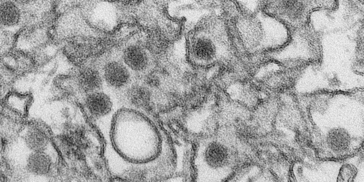
Transmission electron micrograph (TEM) of the Zika virus. Credit: Cynthia Goldsmith/Centers for Disease Control and Prevention
Viruses have limited genetic material – and few proteins – so all the parts have to work very hard. Zika is a great example; the virus only produces 10 proteins. Now, in a study published in the journal PLOS PathogensSanford Burnham Prebys researchers have shown how the virus does so much with so little and may have identified a therapeutic vulnerability.
In the study, the research team showed that Zika’s enzyme, NS2B-NS3, is a versatile tool with two essential functions: breaking down proteins (a protease) and splitting its own double-stranded RNA into single strands ( a helicase).
“We found that the Zika enzyme complex changes function depending on its shape,” says Alexey Terskikh, Ph.D., associate professor at Sanford Burnham Prebys and senior author of the paper. “In the closed conformation, it acts like a classic protease. But then it alternates between open and super-open conformations, which allows it to seize and then release a single strand of RNA – and these functions are essential for viral replication .”
Zika is an RNA virus that is part of a family of deadly pathogens called flaviviruses, which include West Nile, dengue, yellow fever, Japanese encephalitis and others. The virus is transmitted by mosquitoes and infects uterine and placental cells (among other cell types), making it particularly dangerous for pregnant women. Once inside host cells, the virus rearranges them to produce more Zika.
Understanding Zika at the molecular level could have a huge benefit: a therapeutic target. It would be difficult to create safe drugs targeting the domains of the enzyme needed for protease or helicase functions, because human cells have many similar molecules. However, a drug that blocks Zika’s conformational changes could be effective. If the complex cannot change shape, it will not be able to perform its critical functions and no new Zika particles will be produced.
An efficient machine
Researchers have long known that the essential Zika enzyme was made up of two units: NS2B-NS3pro and NS3hel. NS2B-NS3pro performs protease functions, cutting long polypeptides into Zika proteins. However, the abilities of NS2B-NS3pro to bind single-stranded RNA and help separate double-stranded RNA during viral replication have only recently been discovered.
In this study, the researchers built on recent crystal structures and used protein biochemistry, fluorescence polarization, and computational modeling to dissect the life cycle of NS2B-NS3pro. NS3pro is connected to NS3hel (the helicase) by a short amino acid linker and becomes active when the complex is in its closed conformation, like a closed accordion. RNA binding occurs when the complex is open, whereas the complex must go through the super-open conformation to release the RNA.
These conformational changes are driven by the dynamics of NS3hel activity, which extends the linker and ultimately “pulls” on the NS3pro to release the RNA. NS3pro is anchored inside the host cell’s endoplasmic reticulum (ER), a key organelle that helps guide cellular proteins to their proper destinations, via NS2B and, in a closed conformation, cleaves the Zika polypeptide, helping to generate all viral proteins. .
On the other side of the linker, NS3hel separates the Zika double-stranded RNA and conveniently returns one strand to NS3pro, which has positively charged “forks” to hold onto negatively charged RNA.
“There’s a very nice groove of positive charges,” says Terskikh. “So the RNA naturally follows this groove. Then the complex switches to the closed conformation and releases the RNA.”
When NS3hel extends to grab double-stranded RNA, it pulls the complex with it; however, because NS3pro is anchored in the ER membrane and the linker can only extend so far, the complex snaps into the super-open conformation and releases RNA. The complex then relaxes and returns to the open conformation, ready for a new cycle.
Meanwhile, when NS3pro detects a viral polypeptide to be cut, it forces the complex into a closed conformation, thereby becoming a protease. The authors call this process “reverse worming” because the seizure and release of the single-stranded RNA resembles the movements of the worm, but backwards, with the jaws (the protease) trailing behind.
In addition to providing a possible therapeutic target for Zika, this detailed understanding could be applied to other flaviviruses, which share similar molecular machinery.
“Versions of the NS2B-NS3pro complex are found in all flaviviruses,” explains Terskikh. “This could potentially constitute an entirely new class of drug targets for multiple viruses.”
More information:
Sergey A. Shiryaev et al, Dual function of Zika virus NS2B-NS3 protease, PLOS Pathogens (2023). DOI: 10.1371/journal.ppat.1011795
Provided by Sanford-Burnham Prebys
Quote: Study reveals Zika transformation machinery and possible vulnerability (December 9, 2023) retrieved December 10, 2023 from
This document is subject to copyright. Except for fair use for private study or research purposes, no part may be reproduced without written permission. The content is provided for information only.


