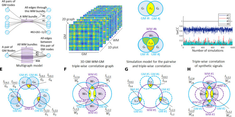
Illustration of the triple correlation network model. Credit: Scientists progress (2024). DOI: 10.1126/sciadv.adi0616
By mapping brain activity in three dimensions, researchers at Vanderbilt University Medical Center have obtained a more detailed picture of how the brain changes with age.
Their findings, described in the January 26 issue of the journal Scientists progressmay help advance the understanding, early diagnosis and treatment of Alzheimer’s disease, bipolar disorder and other disturbances of normal brain function.
Half of the human brain is made up of gray matter, nerve cells that process sensations, control voluntary movements, and enable speech, learning, and cognition. The other half is made up of white matter, the axons that connect areas of gray matter together and project to the rest of the body.
In the field of functional magnetic resonance imaging (fMRI) of the brain, white matter has historically been understudied, in part because it is easier to detect functional signals from gray matter.
Recently, VUMC scientists discovered that they could reliably separate and detect white matter signals, opening the door to a better understanding of the previously neglected other half of the brain.
They have now taken the research to the next level. Through a complex series of mathematical formulas, the paper’s first author, Zhongliang Zu, Ph.D., proposed a method to simultaneously map how areas of gray matter “talk” to each other via brain connections. white matter.
“This is a major expansion of using brain imaging to study brain networks, but including, for the first time, white matter,” said John Gore, Ph.D., director of the Institute for Imaging Sciences at Vanderbilt University and corresponding author of the article. .
Zhongliang Zu, PhD, left, and John Gore, PhD, lead an effort to map brain networks in three dimensions. Credit: Erin O. Smith
A major technique for studying brain activity, fMRI measures changes in blood oxygenation level-dependent (BOLD) signals that, in gray matter, reflect an increase in blood flow (and oxygen) in response to increased neuronal activity.
Although BOLD signals in white matter are less well understood, it is becoming clear that white matter is not a passive tissue that simply connects areas of gray matter.
White matter “plays a central role in human learning processes,” the VUMC scientists say in their article. Changes in white matter microstructure “alter the fidelity of neuronal signal transmissions and, therefore, brain function.”
Previous research has determined functional connectivity between two gray matter areas by correlating their BOLD signals.
As described in the current paper, Zu, a research associate professor of radiology and radiological sciences and biomedical engineering, designed a “triple correlational framework” that integrates white matter signals into the study of functional connectivity pathways.
Using fMRI images of the brains of 490 individuals from publicly available databases and applying multivariate statistical methods, Zu, Gore and their colleagues were able to determine that white matter fibers form multiple and complex gray matter connections .
“A white matter fiber can carry signals from multiple potential inputs to different areas of gray matter,” Gore explained. “At the same time, any pair of gray matter areas can communicate through many different (white matter) pathways.”
Zu’s mathematical equations helped determine how much each pair of gray matter contributes to signal transmission through a single white matter fiber and, at the same time, how much each pair uses different fibers. “It’s really a major milestone,” Gore said.
The integration of white matter signals provides, as the title of the article suggests, the “missing third dimension” for understanding functional connectivity between different brain regions.
By studying brain scans of different age groups, the researchers found that overall connectivity in parts of the brain decreased with age, while brain activation increased in the frontal cortex, involved in higher cognitive functions.
This change may be aimed at offsetting declines in other areas, they noted.
Researchers are currently studying what might be the functional consequences of vascular changes in white matter associated with brain disorders such as Alzheimer’s disease.
In the future, measuring changes in functional connectivity between brain regions could serve as a biomarker, a way to monitor the progression of diseases that affect white matter and response to treatment, Gore said.
More information:
Zhongliang Zu et al, The missing third dimension — Functional correlations of BOLD signals incorporating white matter, Scientists progress (2024). DOI: 10.1126/sciadv.adi0616
Provided by Vanderbilt University
Quote: 3D brain mapping opens a window on the aging brain (February 9, 2024) retrieved February 9, 2024 from
This document is subject to copyright. Apart from fair use for private study or research purposes, no part may be reproduced without written permission. The content is provided for information only.


